当前位置:
X-MOL 学术
›
J. Mater. Chem. B
›
论文详情
Our official English website, www.x-mol.net, welcomes your
feedback! (Note: you will need to create a separate account there.)
Multiplexed fluorescence lifetime imaging by concentration-dependent quenching†
Journal of Materials Chemistry B ( IF 6.1 ) Pub Date : 2018-02-20 00:00:00 , DOI: 10.1039/c8tb00095f Teng Luo 1, 2, 3, 4, 5 , Ting Zhou 1, 2, 3, 4, 5 , Yihua Zhao 1, 2, 3, 4, 5 , Liwei Liu 1, 2, 3, 4, 5 , Junle Qu 1, 2, 3, 4, 5
Journal of Materials Chemistry B ( IF 6.1 ) Pub Date : 2018-02-20 00:00:00 , DOI: 10.1039/c8tb00095f Teng Luo 1, 2, 3, 4, 5 , Ting Zhou 1, 2, 3, 4, 5 , Yihua Zhao 1, 2, 3, 4, 5 , Liwei Liu 1, 2, 3, 4, 5 , Junle Qu 1, 2, 3, 4, 5
Affiliation
|
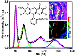
This study sought to use the undesirable concentration-dependent quenching to propose a simple multiplexed imaging analysis for histopathological identification of different stained tissues. To verify this point, the relationship between the fluorescence lifetime and eosin concentration was obtained. At low concentrations, the fluorescence lifetimes of eosin were independent of the concentration (<0.25 μg ml−1). At moderate concentrations (0.25–1 μg ml−1), eosin was quenched and its fluorescence lifetime was shortened gradually. Interestingly, the fluorescence of eosin was still quenched when the concentration exceeded 1 μg ml−1, but its corresponding fluorescence lifetimes increase with increased concentration (>100 μg ml−1). To further verify that multiplexed imaging of different tissues could be achieved only by eosin, we used fluorescence lifetime imaging microscopy (FLIM) to measure fluorescence lifetimes from hematoxylin and eosin (H&E) stained sections. Working directly on an average fluorescence lifetime (τm) histogram for lifetime-based separation easily achieved multiplexed imaging in situ. H&E stained erythrocytes, smooth muscles, collagen and artificial structures on a prepared microscopic slide could be identified without the need of alternating laser excitation, using hyperspectral systems and special staining or multi-labeled immunofluorescence. Using only eosin, different types of tissues could be distinguished by eosin concentration-dependent quenching. Hence, eosin fluorescence lifetimes potentially simplify multiplexed imaging and may have potential applications for pathological diagnosis.
中文翻译:

通过浓度依赖性淬灭进行的多重荧光寿命成像†
这项研究试图使用不良的浓度依赖性猝灭来提出简单的多重成像分析,以用于不同染色组织的组织病理学鉴定。为了证实这一点,获得了荧光寿命和曙红浓度之间的关系。在低浓度下,曙红的荧光寿命与浓度(<0.25μgml -1)无关。在中等浓度(0.25-1μgml -1)下,曙红被淬灭,其荧光寿命逐渐缩短。有趣的是,当浓度超过1μgml -1时,曙红的荧光仍被淬灭,但其相应的荧光寿命随浓度的增加而增加(> 100μgml -1)。为了进一步验证仅曙红可以实现不同组织的多重成像,我们使用了荧光寿命成像显微镜(FLIM)来测量苏木精和曙红(H&E)染色切片的荧光寿命。直接在平均荧光寿命工作(τ米)直方图用于基于寿命分离容易地实现多路成像原位。使用高光谱系统和特殊染色或多标记免疫荧光技术,无需交替进行激光激发,即可在制备的显微镜载玻片上鉴定出H&E染色的红细胞,平滑肌,胶原蛋白和人工结构。仅使用曙红,可以通过曙红浓度依赖性猝灭来区分不同类型的组织。因此,曙红荧光寿命潜在地简化了多重成像,并且可能在病理诊断中具有潜在的应用。
更新日期:2018-02-20
中文翻译:
通过浓度依赖性淬灭进行的多重荧光寿命成像†
这项研究试图使用不良的浓度依赖性猝灭来提出简单的多重成像分析,以用于不同染色组织的组织病理学鉴定。为了证实这一点,获得了荧光寿命和曙红浓度之间的关系。在低浓度下,曙红的荧光寿命与浓度(<0.25μgml -1)无关。在中等浓度(0.25-1μgml -1)下,曙红被淬灭,其荧光寿命逐渐缩短。有趣的是,当浓度超过1μgml -1时,曙红的荧光仍被淬灭,但其相应的荧光寿命随浓度的增加而增加(> 100μgml -1)。为了进一步验证仅曙红可以实现不同组织的多重成像,我们使用了荧光寿命成像显微镜(FLIM)来测量苏木精和曙红(H&E)染色切片的荧光寿命。直接在平均荧光寿命工作(τ米)直方图用于基于寿命分离容易地实现多路成像原位。使用高光谱系统和特殊染色或多标记免疫荧光技术,无需交替进行激光激发,即可在制备的显微镜载玻片上鉴定出H&E染色的红细胞,平滑肌,胶原蛋白和人工结构。仅使用曙红,可以通过曙红浓度依赖性猝灭来区分不同类型的组织。因此,曙红荧光寿命潜在地简化了多重成像,并且可能在病理诊断中具有潜在的应用。


 京公网安备 11010802027423号