Acta Biomaterialia ( IF 9.4 ) Pub Date : 2018-02-17 , DOI: 10.1016/j.actbio.2018.02.012 A.D. Anastasiou , S. Strafford , C.L. Thomson , J. Gardy , T.J. Edwards , M. Malinowski , S.A. Hussain , N.K. Metzger , A. Hassanpour , C.T.A. Brown , A.P. Brown , M.S. Duggal , A. Jha
|
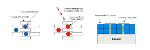
A radical new methodology for the exogenous mineralization of hard tissues is demonstrated in the context of laser-biomaterials interaction. The proposed approach is based on the use of femtosecond pulsed lasers (fs) and Fe3+-doped calcium phosphate minerals (specifically in this work fluorapatite powder containing Fe2O3 nanoparticles (NP)). A layer of the synthetic powder is applied to the surface of eroded bovine enamel and is irradiated with a fs laser (1040 nm wavelength, 1 GHz repetition rate, 150 fs pulse duration and 0.4 W average power). The Fe2O3 NPs absorb the light and may act as thermal antennae, dissipating energy to the vicinal mineral phase. Such a photothermal process triggers the sintering and densification of the surrounding calcium phosphate crystals thereby forming a new, dense layer of typically 20 μm in thickness, which is bonded to the underlying surface of the natural enamel. The dispersed iron oxide NPs, ensure the localization of temperature excursion, minimizing collateral thermal damage to the surrounding natural tissue during laser irradiation. Simulated brushing trials (pH cycle and mechanical force) on the synthetic layer show that the sintered material is more acid resistant than the natural mineral of enamel. Furthermore, nano-indentation confirms that the hardness and Young’s modulus of the new layers are significantly more closely matched to enamel than current restorative materials used in clinical dentistry. Although the results presented herein are exemplified in the context of bovine enamel restoration, the methodology may be more widely applicable to human enamel and other hard-tissue regenerative engineering.
Statement of Significance
In this work we provide a new methodology for the mineralisation of dental hard tissues using femtosecond lasers and iron doped biomaterials. In particular, we demonstrate selective laser sintering of an iron doped fluorapatite on the surface of eroded enamel under low average power and mid-IR wavelength and the formation of a new layer to substitute the removed material. The new layer is evaluated through simulated brushing trials and nano-indentation. From the results we can conclude that is more acid resistant than natural enamel while, its mechanical properties are superior to that of current restorative materials. To the best of our knowledge this is the first time that someone demonstrated, laser sintering and bonding of calcium phosphate biomaterials on hard tissues. Although we here we discuss the case of dental enamel, similar approach can be adopted for other hard tissues, leading to new strategies for the fixation of bone/tooth defects.
中文翻译:
使用光吸收矿物质和飞秒激光对硬组织进行外源性矿化;牙釉质
在激光-生物材料相互作用的背景下,证明了一种用于硬组织外源性矿化的根本方法。提议的方法基于飞秒脉冲激光(fs)和掺Fe 3+的磷酸钙矿物(特别是在这项工作中,含有Fe 2 O 3纳米粒子(NP)的氟磷灰石粉末)的使用。将一层合成粉末涂在侵蚀的牛牙釉质表面,并用fs激光(1040 nm波长,1 GHz重复频率,150 fs脉冲持续时间和0.4 W平均功率)照射。铁2 O 3NP吸收光并可能充当热触角,将能量消散到邻近的矿物质相。这种光热过程触发了周围磷酸钙晶体的烧结和致密化,从而形成了厚度通常为20μm的新的致密层,该层与天然釉质的下层表面粘结在一起。分散的氧化铁纳米颗粒确保了温度偏移的局部性,从而最大程度地减少了激光辐照期间对周围自然组织的附带热损害。在合成层上进行的模拟刷涂试验(pH循环和机械力)表明,该烧结材料比搪瓷的天然矿物更耐酸。此外,纳米压痕证实,新层的硬度和杨氏模量与牙釉质相比,比目前在临床牙科中使用的修复材料更紧密地匹配。尽管本文介绍的结果以牛牙釉质修复为例,但该方法可能更广泛地适用于人类牙釉质和其他硬组织再生工程。
重要声明
在这项工作中,我们提供了使用飞秒激光和掺铁生物材料对牙齿硬组织进行矿化的新方法。特别是,我们演示了在腐蚀的搪瓷表面上对掺铁的氟磷灰石的选择性激光烧结。在低平均功率和中红外波长下形成一层新的层来替代去除的材料。通过模拟刷涂试验和纳米压痕对新层进行评估。从结果可以得出结论,它比天然瓷釉具有更高的耐酸性,同时其机械性能也优于目前的修复材料。据我们所知,这是首次有人证明激光烧结和磷酸钙生物材料在硬组织上的粘结。尽管我们在这里讨论了牙釉质的情况,但其他硬组织也可以采用类似的方法,从而导致了修复骨/齿缺损的新策略。


 京公网安备 11010802027423号