当前位置:
X-MOL 学术
›
Opt. Lett.
›
论文详情
Our official English website, www.x-mol.net, welcomes your
feedback! (Note: you will need to create a separate account there.)
Mid-infrared multispectral tissue imaging using a chalcogenide fiber supercontinuum source
Optics Letters ( IF 3.1 ) Pub Date : 2018-02-21 , DOI: 10.1364/ol.43.000999 Christian Rosenberg Petersen , Nikola Prtljaga , Mark Farries , Jon Ward , Bruce Napier , Gavin Rhys Lloyd , Jayakrupakar Nallala , Nick Stone , Ole Bang
Optics Letters ( IF 3.1 ) Pub Date : 2018-02-21 , DOI: 10.1364/ol.43.000999 Christian Rosenberg Petersen , Nikola Prtljaga , Mark Farries , Jon Ward , Bruce Napier , Gavin Rhys Lloyd , Jayakrupakar Nallala , Nick Stone , Ole Bang
|
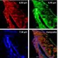
We present, to the best of our knowledge, the first demonstration of mid-infrared supercontinuum (SC) tissue imaging at wavelengths beyond 5 μm using a fiber-coupled SC source spanning 2–7.5 μm. The SC was generated in a tapered large-mode-area chalcogenide photonic crystal fiber in order to obtain broad bandwidth, high average power, and single-mode output for diffraction-limited imaging performance. Tissue imaging was demonstrated in transmission at selected wavelengths between 5.7 () and 7.3 μm () by point scanning over a sub-millimeter region of colon tissue, and the results were compared to images obtained from a commercial instrument.
中文翻译:

使用硫族化物纤维超连续谱源的中红外多光谱组织成像
据我们所知,我们首次展示了使用跨度为2–7.5μm的光纤耦合SC光源对波长超过5μm的中红外超连续谱(SC)组织成像的演示。为了获得宽带宽,高平均功率和用于限制衍射成像性能的单模输出,在锥形大模式面积硫族化物光子晶体光纤中生成了SC。组织成像在5.7()和7.3μm(通过在结肠组织的亚毫米区域上进行点扫描,将结果与从商用仪器获得的图像进行比较。
更新日期:2018-03-01
中文翻译:
使用硫族化物纤维超连续谱源的中红外多光谱组织成像
据我们所知,我们首次展示了使用跨度为2–7.5μm的光纤耦合SC光源对波长超过5μm的中红外超连续谱(SC)组织成像的演示。为了获得宽带宽,高平均功率和用于限制衍射成像性能的单模输出,在锥形大模式面积硫族化物光子晶体光纤中生成了SC。组织成像在5.7()和7.3μm(通过在结肠组织的亚毫米区域上进行点扫描,将结果与从商用仪器获得的图像进行比较。


 京公网安备 11010802027423号