Acta Biomaterialia ( IF 9.4 ) Pub Date : 2018-02-15 , DOI: 10.1016/j.actbio.2018.02.009 Jessica H Brown 1 , Prativa Das 2 , Michael D DiVito 1 , David Ivancic 1 , Lay Poh Tan 3 , Jason A Wertheim 4
|
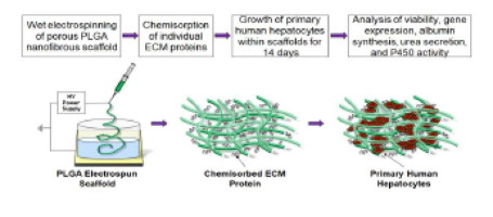
A major challenge of maintaining primary hepatocytes in vitro is progressive loss of hepatocyte-specific functions, such as protein synthesis and cytochrome P450 (CYP450) catalytic activity. We developed a three-dimensional (3D) nanofibrous scaffold made from poly(L-lactide-co-glycolide) (PLGA) polymer using a newly optimized wet electrospinning technique that resulted in a highly porous structure that accommodated inclusion of primary human hepatocytes. Extracellular matrix (ECM) proteins (type I collagen or fibronectin) at varying concentrations were chemically linked to electrospun PLGA using amine coupling to develop an in vitro culture system containing the minimal essential ECM components of the liver micro-environment that preserve hepatocyte function in vitro. Cell-laden nanofiber scaffolds were tested in vitro to maintain hepatocyte function over a two-week period. Incorporation of type I collagen onto PLGA scaffolds (PLGA-Chigh: 100 µg/mL) led to 10-fold greater albumin secretion, 4-fold higher urea synthesis, and elevated transcription of hepatocyte-specific CYP450 genes (CYP3A4, 3.5-fold increase and CYP2C9, 3-fold increase) in primary human hepatocytes compared to the same cells grown within unmodified PLGA scaffolds over two weeks. These indices, measured using collagen-bonded scaffolds, were also higher than scaffolds coupled to fibronectin or an ECM control sandwich culture composed of type I collagen and Matrigel. Induction of CYP2C9 activity was also higher in these same type I collagen PLGA scaffolds compared to other ECM-modified or unmodified PLGA constructs and was equivalent to the ECM control at 7 days. Together, we demonstrate a minimalist ECM-based 3D synthetic scaffold that accommodates primary human hepatocyte inclusion into the matrix, maintains long-term in vitro survival and stimulates function, which can be attributed to coupling of type I collagen.
Statement of Significance
Culturing primary hepatocytes within a three-dimensional (3D) structure that mimics the natural liver environment is a promising strategy for extending the function and viability of hepatocytes in vitro. In the present study we generate porous PLGA nanofibers, that are chemically modified with extracellular matrix proteins, to serve as 3D scaffolds for the in vitro culture of primary human hepatocytes. Our findings demonstrate that the use of ECM proteins, especially type I collagen, in a porous 3D environment helps to improve the synthetic function of primary hepatocytes over time. We believe the work presented within will provide insights to readers for drug toxicity and tissue engineering applications.
中文翻译:
I 型胶原修饰的纳米纤维 PLGA 电纺支架影响肝细胞功能和体外支持活力
体外维持原代肝细胞的一个主要挑战是肝细胞特异性功能的逐渐丧失,例如蛋白质合成和细胞色素 P450 (CYP450) 催化活性。我们使用新优化的湿式静电纺丝技术开发了一种由聚(L-丙交酯-共-乙交酯)(PLGA)聚合物制成的三维(3D)纳米纤维支架,该支架形成高度多孔的结构,可容纳原代人肝细胞。使用胺偶联将不同浓度的细胞外基质 (ECM) 蛋白(I 型胶原蛋白或纤连蛋白)与电纺 PLGA 化学连接,以开发体外培养系统,该系统包含肝脏微环境的最低必需 ECM 成分,可在体外保留肝细胞功能。体外测试了充满细胞的纳米纤维支架在两周内维持肝细胞功能的情况。将 I 型胶原蛋白掺入 PLGA 支架上(PLGA-C高:100 µg/mL)导致白蛋白分泌增加 10 倍,尿素合成增加 4 倍,肝细胞特异性 CYP450 基因(CYP3A4,3.5 倍)的转录增加与在未修饰的 PLGA 支架中生长超过两周的相同细胞相比,原代人肝细胞中的 CYP2C9 增加和 CYP2C9 增加 3 倍)。使用胶原蛋白粘合支架测量的这些指数也高于与纤连蛋白偶联的支架或由 I 型胶原蛋白和基质胶组成的 ECM 对照夹心培养物的支架。与其他 ECM 修饰或未修饰的 PLGA 构建体相比,这些相同的 I 型胶原 PLGA 支架中 CYP2C9 活性的诱导也更高,并且在 7 天时与 ECM 对照相当。我们共同展示了一种基于 ECM 的极简 3D 合成支架,可将原代人肝细胞容纳到基质中,维持长期体外存活并刺激功能,这可归因于 I 型胶原蛋白的偶联。
重要性声明
在模拟自然肝脏环境的三维 (3D) 结构中培养原代肝细胞是在体外扩展肝细胞功能和活力的一种有前途的策略。在本研究中,我们生成多孔 PLGA 纳米纤维,并用细胞外基质蛋白进行化学修饰,作为原代人肝细胞体外培养的 3D 支架。我们的研究结果表明,在多孔 3D 环境中使用 ECM 蛋白,尤其是 I 型胶原蛋白,有助于随着时间的推移改善原代肝细胞的合成功能。我们相信,其中介绍的工作将为读者提供有关药物毒性和组织工程应用的见解。


 京公网安备 11010802027423号