当前位置:
X-MOL 学术
›
Chem. Sci.
›
论文详情
Our official English website, www.x-mol.net, welcomes your
feedback! (Note: you will need to create a separate account there.)
Highly multiplexed single-cell in situ RNA and DNA analysis with bioorthogonal cleavable fluorescent oligonucleotides†
Chemical Science ( IF 7.6 ) Pub Date : 2018-02-13 00:00:00 , DOI: 10.1039/c7sc05089e Manas Mondal 1 , Renjie Liao 1 , Christopher D Nazaroff 1, 2 , Adam D Samuel 1 , Jia Guo 1
Chemical Science ( IF 7.6 ) Pub Date : 2018-02-13 00:00:00 , DOI: 10.1039/c7sc05089e Manas Mondal 1 , Renjie Liao 1 , Christopher D Nazaroff 1, 2 , Adam D Samuel 1 , Jia Guo 1
Affiliation
|
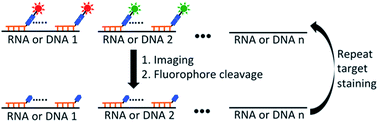
The ability to profile transcripts and genomic loci comprehensively in single cells in situ is essential to advance our understanding of normal physiology and disease pathogenesis. Here we report a highly multiplexed single-cell in situ RNA and DNA analysis approach using bioorthogonal cleavable fluorescent oligonucleotides. In this approach, oligonucleotides tethered to fluorophores through an azide-based cleavable linker are used to detect their nucleic acids targets by in situ hybridization. After fluorescence imaging, the fluorophores in the whole specimen are efficiently cleaved in 30 minutes without loss of RNA or DNA integrity. Through reiterative cycles of hybridization, imaging, and cleavage, this method has the potential to quantify hundreds to thousands of different RNA species or genomic loci in single cells in situ at the single-molecule sensitivity. Applying this approach, we demonstrate that different nucleic acids can be detected in each hybridization cycle by multi-color staining, and at least ten continuous hybridization cycles can be carried out in the same specimen. We also show that the integrated single-cell in situ analysis of DNA, RNA and protein can be achieved using cleavable fluorescent oligonucleotides combined with cleavable fluorescent antibodies. This highly multiplexed imaging platform will have wide applications in systems biology and biomedical research.
中文翻译:

使用生物正交可切割荧光寡核苷酸进行高度多重单细胞原位 RNA 和 DNA 分析†
原位全面分析单细胞转录本和基因组位点的能力对于增进我们对正常生理学和疾病发病机制的理解至关重要。在这里,我们报告了一种使用生物正交可切割荧光寡核苷酸的高度多重单细胞原位RNA 和 DNA 分析方法。在这种方法中,寡核苷酸通过基于叠氮化物的可切割接头连接到荧光团,用于通过原位杂交检测其核酸靶标。荧光成像后,整个样本中的荧光团在 30 分钟内被有效切割,而不会损失 RNA 或 DNA 的完整性。通过重复的杂交、成像和切割循环,该方法有可能以单分子灵敏度原位定量单细胞中数百至数千个不同的 RNA 种类或基因组位点。应用这种方法,我们证明可以通过多色染色在每个杂交循环中检测到不同的核酸,并且可以在同一样本中进行至少十个连续杂交循环。我们还表明,使用可裂解荧光寡核苷酸与可裂解荧光抗体相结合,可以实现 DNA、RNA 和蛋白质的集成单细胞原位分析。这种高度多重成像平台将在系统生物学和生物医学研究中具有广泛的应用。
更新日期:2018-02-13
中文翻译:
使用生物正交可切割荧光寡核苷酸进行高度多重单细胞原位 RNA 和 DNA 分析†
原位全面分析单细胞转录本和基因组位点的能力对于增进我们对正常生理学和疾病发病机制的理解至关重要。在这里,我们报告了一种使用生物正交可切割荧光寡核苷酸的高度多重单细胞原位RNA 和 DNA 分析方法。在这种方法中,寡核苷酸通过基于叠氮化物的可切割接头连接到荧光团,用于通过原位杂交检测其核酸靶标。荧光成像后,整个样本中的荧光团在 30 分钟内被有效切割,而不会损失 RNA 或 DNA 的完整性。通过重复的杂交、成像和切割循环,该方法有可能以单分子灵敏度原位定量单细胞中数百至数千个不同的 RNA 种类或基因组位点。应用这种方法,我们证明可以通过多色染色在每个杂交循环中检测到不同的核酸,并且可以在同一样本中进行至少十个连续杂交循环。我们还表明,使用可裂解荧光寡核苷酸与可裂解荧光抗体相结合,可以实现 DNA、RNA 和蛋白质的集成单细胞原位分析。这种高度多重成像平台将在系统生物学和生物医学研究中具有广泛的应用。


 京公网安备 11010802027423号