当前位置:
X-MOL 学术
›
Chem. Sci.
›
论文详情
Our official English website, www.x-mol.net, welcomes your
feedback! (Note: you will need to create a separate account there.)
Nanobubble-embedded inorganic 808 nm excited upconversion nanocomposites for tumor multiple imaging and treatment†
Chemical Science ( IF 7.6 ) Pub Date : 2018-02-09 00:00:00 , DOI: 10.1039/c8sc00108a Ming-Hsien Chan,Yu-Ting Pan,Yung-Chieh Chan,Michael Hsiao,Chung-Hsuan Chen,Lingdong Sun,Ru-Shi Liu
Chemical Science ( IF 7.6 ) Pub Date : 2018-02-09 00:00:00 , DOI: 10.1039/c8sc00108a Ming-Hsien Chan,Yu-Ting Pan,Yung-Chieh Chan,Michael Hsiao,Chung-Hsuan Chen,Lingdong Sun,Ru-Shi Liu
|
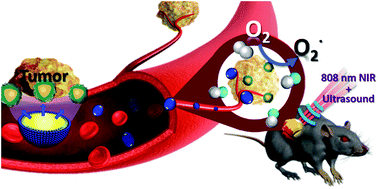
Contrast reagents for ultrasound imaging are widely used in clinical medical diagnosis because ultrasound resolution is limited. Contrast agents must be utilized to enhance the image resolution. At present, microbubbles (MBs) and nanobubbles (NBs) are the main contrast reagent candidates for improving the signal resolution. Fluorescence upconversion nanoparticles provide high sensitivity and also function as nanocarriers. This can label tumor cells in a specific organ under irradiation of near-infrared (NIR) light. However, despite the use of NIR light, the penetration depth of NIR is only approximately 15 mm. Thus, we combine fluorescence with ultrasonic imaging to achieve the effect of multiple imaging and solve the low penetration depth of fluorescence imaging and the poor resolution of ultrasound imaging. The dual imaging modalities achieved higher resolution or signal to noise ratios. In this study, Nd3+-sensitized upconversion nanoparticles (UCNPs) are combined with graphitic carbon nitride quantum dots (CNs) and embedded in NBs (UCNP–CN@NBs). The UCNPs are excited by 808 nm light and emit visible and ultraviolet light. Then, the energy of the ultraviolet light is transferred to the CNs to produce reactive oxygen species (ROS) for photodynamic therapy. Ultrasonic waves are also used to promote NB bursting and the release of ROS molecules in photodynamic therapy, leading to cancer cell apoptosis.
中文翻译:

用于肿瘤多重成像和治疗的纳米气泡嵌入无机 808 nm 激发上转换纳米复合材料†
由于超声分辨率有限,用于超声成像的造影剂被广泛用于临床医学诊断。必须使用造影剂来提高图像分辨率。目前,微泡(MBs)和纳米泡(NBs)是提高信号分辨率的主要对比剂候选物。荧光上转换纳米粒子提供高灵敏度并且还用作纳米载体。这可以在近红外 (NIR) 光的照射下标记特定器官中的肿瘤细胞。然而,尽管使用了近红外光,近红外光的穿透深度只有大约 15 毫米。因此,我们将荧光与超声成像相结合,达到多次成像的效果,解决了荧光成像穿透深度低、超声成像分辨率差的问题。双成像模式实现了更高的分辨率或信噪比。在本研究中,Nd3+敏化上转换纳米粒子 (UCNPs) 与石墨氮化碳量子点 (CNs) 结合并嵌入到 NBs (UCNP-CN@NBs) 中。UCNPs 被 808 nm 光激发并发射可见光和紫外光。然后,紫外光的能量被转移到 CNs 以产生活性氧 (ROS) 用于光动力治疗。在光动力治疗中,超声波还用于促进 NB 爆发和 ROS 分子的释放,导致癌细胞凋亡。
更新日期:2018-02-09
中文翻译:
用于肿瘤多重成像和治疗的纳米气泡嵌入无机 808 nm 激发上转换纳米复合材料†
由于超声分辨率有限,用于超声成像的造影剂被广泛用于临床医学诊断。必须使用造影剂来提高图像分辨率。目前,微泡(MBs)和纳米泡(NBs)是提高信号分辨率的主要对比剂候选物。荧光上转换纳米粒子提供高灵敏度并且还用作纳米载体。这可以在近红外 (NIR) 光的照射下标记特定器官中的肿瘤细胞。然而,尽管使用了近红外光,近红外光的穿透深度只有大约 15 毫米。因此,我们将荧光与超声成像相结合,达到多次成像的效果,解决了荧光成像穿透深度低、超声成像分辨率差的问题。双成像模式实现了更高的分辨率或信噪比。在本研究中,Nd3+敏化上转换纳米粒子 (UCNPs) 与石墨氮化碳量子点 (CNs) 结合并嵌入到 NBs (UCNP-CN@NBs) 中。UCNPs 被 808 nm 光激发并发射可见光和紫外光。然后,紫外光的能量被转移到 CNs 以产生活性氧 (ROS) 用于光动力治疗。在光动力治疗中,超声波还用于促进 NB 爆发和 ROS 分子的释放,导致癌细胞凋亡。


 京公网安备 11010802027423号