当前位置:
X-MOL 学术
›
J. Mater. Chem. B
›
论文详情
Our official English website, www.x-mol.net, welcomes your
feedback! (Note: you will need to create a separate account there.)
Magnetic resonance imaging quantification and biodistribution of magnetic nanoparticles using T1-enhanced contrast†
Journal of Materials Chemistry B ( IF 6.1 ) Pub Date : 2018-01-26 00:00:00 , DOI: 10.1039/c7tb03129g Y. B. Lv 1, 2, 3, 4 , P. Chandrasekharan 3, 4, 5, 6, 7 , Y. Li 8, 9, 10, 11, 12 , X. L. Liu 13, 14, 15, 16 , J. P. Avila 3, 4, 5, 6 , Y. Yang 1, 2, 3, 4 , K. H. Chuang 3, 4, 5, 6, 7 , Xing-Jie Liang 13, 14, 15, 16 , J. Ding 1, 2, 3, 4
Journal of Materials Chemistry B ( IF 6.1 ) Pub Date : 2018-01-26 00:00:00 , DOI: 10.1039/c7tb03129g Y. B. Lv 1, 2, 3, 4 , P. Chandrasekharan 3, 4, 5, 6, 7 , Y. Li 8, 9, 10, 11, 12 , X. L. Liu 13, 14, 15, 16 , J. P. Avila 3, 4, 5, 6 , Y. Yang 1, 2, 3, 4 , K. H. Chuang 3, 4, 5, 6, 7 , Xing-Jie Liang 13, 14, 15, 16 , J. Ding 1, 2, 3, 4
Affiliation
|
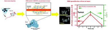
Magnetic iron oxide nanoparticles have been used for various applications such as in the treatment of iron deficiency, as theranostic agents, and as drug carriers. The effective delivery of magnetic iron oxide nanoparticles into the lesion and iron quantification are vital for in vivo theranostic application. To determine the feasibility of using T1 contrast to non-invasively quantify and monitor the IONPs in vivo, monodispersed Gd-doped iron oxide nanoparticles (GdIONPs) with 4 nm core size were fabricated and were used as T1-weighted contrast agents to quantify iron contents based on MRI longitudinal relaxation times (T1). Signal enhancement in positive T1 contrast caused by GdIONPs was observed in this work. The in vivo T1 relaxivity of GdIONPs in a tumor matched well with both in vitro T1 relaxivity and ICP-MS results, demonstrating that the concentration of iron at the tumor site can be directly read from real-time in vivo MRI T1 relaxivity. Hence, by using this strategy, the Fe content in the lesion can be accurately monitored based on MRI longitudinal relaxation times, and this may shed light on effective magnetic hyperthermia cancer therapy in future.
中文翻译:

使用T 1增强的对比度对磁性纳米粒子的磁共振成像定量和生物分布†
磁性氧化铁纳米粒子已用于多种应用,例如治疗铁缺乏症,治疗治疗剂和药物载体。将磁性氧化铁纳米颗粒有效地递送到病变中和铁定量对于体内治疗疗法的应用是至关重要的。为了确定使用T 1造影剂进行体内无创定量和监测IONP的可行性,制备了核芯尺寸为4 nm的单分散Gd掺杂氧化铁纳米颗粒(GdIONP),并用作T 1加权造影剂进行定量铁含量基于MRI的纵向弛豫时间(T 1)。正T信号增强在这项工作中观察到1由GdIONPs引起的对比。的体内T 1与两个匹配良好肿瘤GdIONPs的弛豫在体外诱生T 1个弛豫和ICP-MS的结果,表明铁的在肿瘤部位的浓度可以是直接从实时读体内MRI Ť 1个弛豫。因此,通过使用这种策略,可以基于MRI的纵向弛豫时间准确地监测病变中的Fe含量,这可能为将来有效的磁热疗癌症治疗提供参考。
更新日期:2018-01-26
中文翻译:
使用T 1增强的对比度对磁性纳米粒子的磁共振成像定量和生物分布†
磁性氧化铁纳米粒子已用于多种应用,例如治疗铁缺乏症,治疗治疗剂和药物载体。将磁性氧化铁纳米颗粒有效地递送到病变中和铁定量对于体内治疗疗法的应用是至关重要的。为了确定使用T 1造影剂进行体内无创定量和监测IONP的可行性,制备了核芯尺寸为4 nm的单分散Gd掺杂氧化铁纳米颗粒(GdIONP),并用作T 1加权造影剂进行定量铁含量基于MRI的纵向弛豫时间(T 1)。正T信号增强在这项工作中观察到1由GdIONPs引起的对比。的体内T 1与两个匹配良好肿瘤GdIONPs的弛豫在体外诱生T 1个弛豫和ICP-MS的结果,表明铁的在肿瘤部位的浓度可以是直接从实时读体内MRI Ť 1个弛豫。因此,通过使用这种策略,可以基于MRI的纵向弛豫时间准确地监测病变中的Fe含量,这可能为将来有效的磁热疗癌症治疗提供参考。


 京公网安备 11010802027423号