PLOS ONE ( IF 2.9 ) Pub Date : 2018-01-16 , DOI: 10.1371/journal.pone.0191306 Naoki Okumura , Daiki Matsumoto , Yuya Fukui , Masataka Teramoto , Hirofumi Imai , Tetta Kurosawa , Tomoki Shimada , Friedrich Kruse , Ursula Schlötzer-Schrehardt , Shigeru Kinoshita , Noriko Koizumi
|
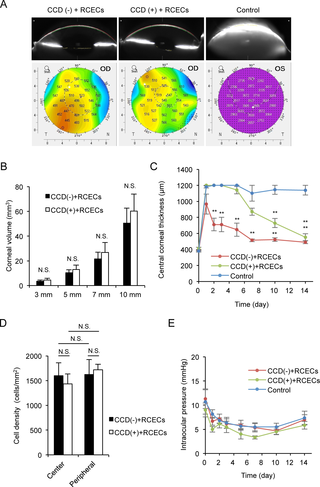
Corneal transparency is maintained by the corneal endothelium through its pump and barrier function. Severe corneal endothelial damage results in dysregulation of water flow and eventually causes corneal haziness and deterioration of visual function. In 2013, we initiated clinical research of cell-based therapy for treating corneal decompensation. In that study, we removed an 8-mm diameter section of damaged corneal endothelium without removing Descemet’s membrane (the basement membrane of the corneal endothelium) and then injected cultured human corneal endothelial cells (CECs) into the anterior chamber. However, Descemet’s membrane exhibits clinically abnormal structural features [i.e., multiple collagenous excrescences (guttae) and thickening] in patients with Fuchs endothelial corneal dystrophy (FECD) and the advanced cornea guttae adversely affects the quality of vision, even in patients without corneal edema. The turnover time of cornea guttae is also not certain. Therefore, we used a rabbit model to evaluate the feasibility of Descemet’s membrane removal in the optical zone only, by performing a small 4-mm diameter descemetorhexis prior to CEC injection. We showed that the corneal endothelium is regenerated both on the corneal stroma (the area of Descemet’s membrane removal) and on the intact peripheral Descemet’s membrane, based on the expression of function-related markers and the restoration of corneal transparency. Recovery of the corneal transparency and central corneal thickness was delayed in areas of Descemet’s membrane removal, but the cell density of the regenerated corneal endothelium and the thickness of the central corneal did not differ between the areas with and without residual Descemet’s membrane at 14 days after CEC injection. Here, we demonstrate that removal of a pathological Descemet’s membrane by a small descemetorhexis is a feasible procedure for use in combination with cell-based therapy. The current strategy might be beneficial for improving visual quality after CEC injection as a treatment for FECD.
中文翻译:
基于细胞的疗法联合地塞米松治疗兔模型的Fuchs内皮角膜营养不良的可行性
角膜内皮通过其泵和屏障功能维持角膜透明性。严重的角膜内皮损伤导致水流失调,并最终导致角膜混浊和视觉功能下降。2013年,我们启动了基于细胞的疗法治疗角膜代偿失调的临床研究。在该研究中,我们去除了受损的角膜内皮的直径为8毫米的部分,而没有去除Descemet的膜(角膜内皮的基底膜),然后将培养的人角膜内皮细胞(CEC)注入前房。但是,Descemet的膜表现出临床异常的结构特征[即,Fuchs内皮角膜营养不良(FECD)和晚期角膜胶质患者的多次胶原性脱落(牙龈炎和增厚)甚至会对视力质量产生不利影响,即使在没有角膜浮肿的患者中也是如此。角膜胶的周转时间也不确定。因此,我们使用兔子模型通过在CEC注射之前进行小直径4mm的去角膜置换术来评估仅在光学区域除去Descemet膜的可行性。我们显示,基于功能相关标记物的表达和角膜透明性的恢复,角膜内皮在角膜基质(Descemet膜去除的区域)和完整的外周Descemet膜上均可再生。Descemet膜去除区域的角膜透明性和中央角膜厚度的恢复被延迟,但是在有和没有残余Descemet膜的区域之间,再生的角膜内皮细胞密度和中央角膜厚度在14天后没有差异。 CEC注射。在这里,我们证明了通过小的去角膜切除术去除病理性Descemet膜是与基于细胞的治疗联合使用的可行方法。当前的策略可能对改善CEC注射后的视觉质量(作为FECD的治疗方法)有益。我们证明,通过小去角膜切除术去除病理性Descemet膜是与基于细胞的治疗联合使用的可行方法。当前的策略可能对改善CEC注射后的视觉质量(作为FECD的治疗方法)有益。我们证明,通过小去角膜切除术去除病理性Descemet膜是与基于细胞的治疗联合使用的可行方法。当前的策略可能对改善CEC注射后的视觉质量(作为FECD的治疗方法)有益。


 京公网安备 11010802027423号