当前位置:
X-MOL 学术
›
ACS Biomater. Sci. Eng.
›
论文详情
Our official English website, www.x-mol.net, welcomes your
feedback! (Note: you will need to create a separate account there.)
Bioinspired Mineralization Using Chondrocyte Membrane Nanofragments
ACS Biomaterials Science & Engineering ( IF 5.4 ) Pub Date : 2018-01-16 00:00:00 , DOI: 10.1021/acsbiomaterials.7b00962 Emilio Satoshi Hara , Masahiro Okada , Noriyuki Nagaoka , Takako Hattori , Takuo Kuboki , Takayoshi Nakano 1 , Takuya Matsumoto
ACS Biomaterials Science & Engineering ( IF 5.4 ) Pub Date : 2018-01-16 00:00:00 , DOI: 10.1021/acsbiomaterials.7b00962 Emilio Satoshi Hara , Masahiro Okada , Noriyuki Nagaoka , Takako Hattori , Takuo Kuboki , Takayoshi Nakano 1 , Takuya Matsumoto
Affiliation

|
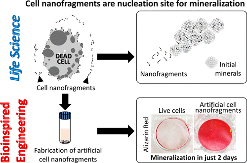
Biomineralization involves complex processes and interactions between organic and inorganic matters, which are controlled in part by the cells. The objectives of this study were, first, to perform a systematic and ultrastructural investigation of the initial mineral formation during secondary ossification center of mouse femur based on material science and biology viewpoint, and then develop novel biomaterials for mineralization based on the in vivo findings. First, we identified the very initial mineral deposition at postnatal day 5 (P5) at the medial side of femur epiphysis by nanocomputed tomography. Initial minerals were found in the surroundings of hypertrophic chondrocytes. Interestingly, histological and immunohistochemical analyses showed that initial mineralization until P6 was based on chondrocyte activity only, i.e., it occurred in the absence of osteoblasts. Moreover, electron microscopy-based ultrastructural analysis showed that cell-secreted matrix vesicles were absent in the early steps of osteoblast-independent endochondral ossification. Instead, chondrocyte membrane nanofragments were found in the fibrous matrix surrounding the hypertrophic chondrocytes. EDS analysis and electron diffraction study indicated that cell membrane nanofragments were not mineralized material, and could be the nucleation site for the newly formed calcospherites. The phospholipids in the cell membrane nanofragments could be a source of phosphate for subsequent calcium phosphate formation, which initially was amorphous, and later transformed into apatite crystals. Finally, artificial cell nanofragments were synthesized from ATDC5 chondrogenic cells, and in vitro assays showed that these nanofragments could promote mineral formation. Taken together, these results indicated that cell membrane nanofragments were the nucleation site for mineral formation, and could potentially be used as material for manipulation of biomineralization.
中文翻译:

利用软骨细胞膜纳米片段的生物启发矿化作用
生物矿化涉及复杂的过程以及有机物和无机物之间的相互作用,而有机物和无机物部分地受细胞控制。这项研究的目的是,首先,基于材料科学和生物学的观点,对小鼠股骨继发骨化中心的初始矿物质形成进行系统的超微结构研究,然后根据体内研究结果开发用于矿化的新型生物材料。首先,我们通过纳米计算机断层扫描技术确定了在产后第5天(P5)在股骨骨physi内侧的最初矿物质沉积。在肥大的软骨细胞周围发现了最初的矿物质。有趣的是,组织学和免疫组织化学分析表明直到P6的最初矿化仅基于软骨细胞的活性,即 它发生在没有成骨细胞的情况下。此外,基于电子显微镜的超微结构分析表明,在成骨细胞独立的软骨内骨化的早期阶段不存在细胞分泌的基质囊泡。相反,在肥大软骨细胞周围的纤维基质中发现了软骨细胞膜纳米片段。EDS分析和电子衍射研究表明,细胞膜纳米碎片不是矿化物质,可能是新形成的钙绿石的成核位点。细胞膜纳米片段中的磷脂可能是随后形成磷酸钙的磷酸盐来源,磷酸钙最初是无定形的,后来转变为磷灰石晶体。最后,从ATDC5软骨细胞合成了人工细胞纳米片段,体外试验表明,这些纳米片段可以促进矿物质的形成。综上所述,这些结果表明细胞膜纳米片段是矿物质形成的成核位点,并有可能被用作生物矿化的处理材料。
更新日期:2018-01-16
中文翻译:

利用软骨细胞膜纳米片段的生物启发矿化作用
生物矿化涉及复杂的过程以及有机物和无机物之间的相互作用,而有机物和无机物部分地受细胞控制。这项研究的目的是,首先,基于材料科学和生物学的观点,对小鼠股骨继发骨化中心的初始矿物质形成进行系统的超微结构研究,然后根据体内研究结果开发用于矿化的新型生物材料。首先,我们通过纳米计算机断层扫描技术确定了在产后第5天(P5)在股骨骨physi内侧的最初矿物质沉积。在肥大的软骨细胞周围发现了最初的矿物质。有趣的是,组织学和免疫组织化学分析表明直到P6的最初矿化仅基于软骨细胞的活性,即 它发生在没有成骨细胞的情况下。此外,基于电子显微镜的超微结构分析表明,在成骨细胞独立的软骨内骨化的早期阶段不存在细胞分泌的基质囊泡。相反,在肥大软骨细胞周围的纤维基质中发现了软骨细胞膜纳米片段。EDS分析和电子衍射研究表明,细胞膜纳米碎片不是矿化物质,可能是新形成的钙绿石的成核位点。细胞膜纳米片段中的磷脂可能是随后形成磷酸钙的磷酸盐来源,磷酸钙最初是无定形的,后来转变为磷灰石晶体。最后,从ATDC5软骨细胞合成了人工细胞纳米片段,体外试验表明,这些纳米片段可以促进矿物质的形成。综上所述,这些结果表明细胞膜纳米片段是矿物质形成的成核位点,并有可能被用作生物矿化的处理材料。


 京公网安备 11010802027423号