当前位置:
X-MOL 学术
›
Eur. Polym. J.
›
论文详情
Our official English website, www.x-mol.net, welcomes your
feedback! (Note: you will need to create a separate account there.)
Structural characterization of nanoparticles formed by fluorinated poly(2-oxazoline)-based polyphiles
European Polymer Journal ( IF 5.8 ) Pub Date : 2018-02-01 , DOI: 10.1016/j.eurpolymj.2018.01.007 Anna Riabtseva , Leonid I. Kaberov , Laurence Noirez , Vasyl Ryukhtin , Corinne Nardin , Bart Verbraeken , Richard Hoogenboom , Petr Stepanek , Sergey K. Filippov
European Polymer Journal ( IF 5.8 ) Pub Date : 2018-02-01 , DOI: 10.1016/j.eurpolymj.2018.01.007 Anna Riabtseva , Leonid I. Kaberov , Laurence Noirez , Vasyl Ryukhtin , Corinne Nardin , Bart Verbraeken , Richard Hoogenboom , Petr Stepanek , Sergey K. Filippov
|
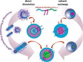
Abstract We report on the self-assembly behavior of poly(2-methyl-2-oxazoline)–block–poly(2-octyl-2-oxazoline) comprising different terminal perfluoroalkyl fragments in aqueous solutions. As reported previously [Kaberov et al. (2017)] such polyphiles can form a plethora of nanostructures depending of the composition and on the way of preparation. Here we report, for the first time, detailed information on the internal structure of the nanoparticles resulting from the self-assembly of these copolymers. Small-angle neutron and X-ray scattering (SANS/SAXS) experiments unambiguously prove the existence of polymersomes, wormlike micelles and their aggregates in aqueous solution. It is shown that increasing content of fluorine in the poly(2-oxazoline) copolymers results in a morphological transition from bilayered or multi-layered vesicles to wormlike micelles for solutions prepared by direct dissolution. In contrast, nanoparticles prepared by dialysis of a polymer solution in a non-selective organic solvent against water are characterized by SAXS method. The internal structure of the nanoparticles could be assessed by fitting of the scattering data, revealing complex core-double shell architecture of spherical symmetry. Additionally, long range ordering is identified for all studied nanoparticles due to the crystallization of the poly(2-octyl-2-oxazoline) segments inside the nanoparticles.
中文翻译:

由氟化聚(2-恶唑啉)基多亲体形成的纳米颗粒的结构表征
摘要 我们报告了包含不同末端全氟烷基片段的聚(2-甲基-2-恶唑啉)-嵌段-聚(2-辛基-2-恶唑啉)在水溶液中的自组装行为。如前所述 [Kabrov et al. (2017)] 根据组成和制备方式,这种多亲体可以形成过多的纳米结构。在这里,我们首次报告了这些共聚物自组装产生的纳米粒子内部结构的详细信息。小角度中子和 X 射线散射 (SANS/SAXS) 实验明确证明了聚合物囊泡、蠕虫状胶束及其在水溶液中的聚集体的存在。结果表明,聚(2-恶唑啉)共聚物中氟含量的增加导致直接溶解制备的溶液从双层或多层囊泡到蠕虫状胶束的形态转变。相比之下,通过在非选择性有机溶剂中对水透析聚合物溶液制备的纳米颗粒通过 SAXS 方法进行表征。纳米粒子的内部结构可以通过拟合散射数据来评估,揭示球对称的复杂核双壳结构。此外,由于纳米颗粒内聚(2-辛基-2-恶唑啉)链段的结晶,所有研究的纳米颗粒都确定了长程排序。通过在对水的非选择性有机溶剂中透析聚合物溶液制备的纳米颗粒通过 SAXS 方法进行表征。纳米粒子的内部结构可以通过拟合散射数据来评估,揭示球对称的复杂核双壳结构。此外,由于纳米颗粒内聚(2-辛基-2-恶唑啉)链段的结晶,所有研究的纳米颗粒都确定了长程排序。通过在对水的非选择性有机溶剂中透析聚合物溶液制备的纳米颗粒通过 SAXS 方法进行表征。纳米粒子的内部结构可以通过拟合散射数据来评估,揭示球对称的复杂核双壳结构。此外,由于纳米颗粒内聚(2-辛基-2-恶唑啉)链段的结晶,所有研究的纳米颗粒都确定了长程排序。
更新日期:2018-02-01
中文翻译:
由氟化聚(2-恶唑啉)基多亲体形成的纳米颗粒的结构表征
摘要 我们报告了包含不同末端全氟烷基片段的聚(2-甲基-2-恶唑啉)-嵌段-聚(2-辛基-2-恶唑啉)在水溶液中的自组装行为。如前所述 [Kabrov et al. (2017)] 根据组成和制备方式,这种多亲体可以形成过多的纳米结构。在这里,我们首次报告了这些共聚物自组装产生的纳米粒子内部结构的详细信息。小角度中子和 X 射线散射 (SANS/SAXS) 实验明确证明了聚合物囊泡、蠕虫状胶束及其在水溶液中的聚集体的存在。结果表明,聚(2-恶唑啉)共聚物中氟含量的增加导致直接溶解制备的溶液从双层或多层囊泡到蠕虫状胶束的形态转变。相比之下,通过在非选择性有机溶剂中对水透析聚合物溶液制备的纳米颗粒通过 SAXS 方法进行表征。纳米粒子的内部结构可以通过拟合散射数据来评估,揭示球对称的复杂核双壳结构。此外,由于纳米颗粒内聚(2-辛基-2-恶唑啉)链段的结晶,所有研究的纳米颗粒都确定了长程排序。通过在对水的非选择性有机溶剂中透析聚合物溶液制备的纳米颗粒通过 SAXS 方法进行表征。纳米粒子的内部结构可以通过拟合散射数据来评估,揭示球对称的复杂核双壳结构。此外,由于纳米颗粒内聚(2-辛基-2-恶唑啉)链段的结晶,所有研究的纳米颗粒都确定了长程排序。通过在对水的非选择性有机溶剂中透析聚合物溶液制备的纳米颗粒通过 SAXS 方法进行表征。纳米粒子的内部结构可以通过拟合散射数据来评估,揭示球对称的复杂核双壳结构。此外,由于纳米颗粒内聚(2-辛基-2-恶唑啉)链段的结晶,所有研究的纳米颗粒都确定了长程排序。


 京公网安备 11010802027423号