当前位置:
X-MOL 学术
›
Biomater. Sci.
›
论文详情
Our official English website, www.x-mol.net, welcomes your feedback! (Note: you will need to create a separate account there.)
Enhanced bone defect repairing effects in glucocorticoid-induced osteonecrosis of the femoral head using a porous nano-lithium-hydroxyapatite/gelatin microsphere/erythropoietin composite scaffold†
Biomaterials Science ( IF 6.6 ) Pub Date : 2018-01-10 00:00:00 , DOI: 10.1039/c7bm00975e Donghai Li 1 , Xiaowei Xie , Zhouyuan Yang , Changde Wang , Zhun Wei , Pengde Kang
Biomaterials Science ( IF 6.6 ) Pub Date : 2018-01-10 00:00:00 , DOI: 10.1039/c7bm00975e Donghai Li 1 , Xiaowei Xie , Zhouyuan Yang , Changde Wang , Zhun Wei , Pengde Kang
Affiliation

|
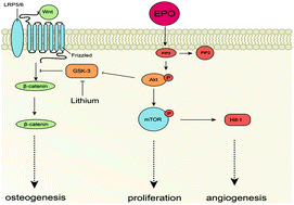
Glucocorticoid-induced osteonecrosis of the femoral head (GIONFH) is a common debilitating disease that occurs in young and middle-aged adults. To treat early GIONFH, core decompression and bone graft are regarded as effective measures. However, the ideal bone graft should possess bioactivity as well as biomechanical properties. The most commonly used bone graft materials are currently unsatisfactory. In this study, we fabricated a composited scaffold using lithium (Li) to activate the Wnt signal pathway and erythrogenin (EPO) to upregulate the HIF-1/VEGF pathway to improve the osteogenic and angiogenic effects of the scaffold. We obtained the porous gelatin/nano-lithium-hydroxyapatite/gelatin microsphere/rhEPO (Li-nHA/GMs/rhEPO) composited scaffold and assessed its mechanical properties, release properties, and in vitro bioactivity. Then, we implanted the scaffold into the femoral heads of GIONFH rabbits after core decompression surgery and evaluated the osteogenic and angiogenic abilities of the scaffold in vivo as well as its bone defect repair efficacy. As the results show, the Li-nHA/GM/rhEPO scaffold possessed good mechanical compression strength and enabled continuous release of Li and rhEPO. Moreover, the scaffold improved the viability of glucocorticoid-treated BMMSCs and vascular endothelial cells and increased the expression of osteogenic and angiogenic factors. In the in vivo study, the composited scaffold improved new bone formation and exerted effects on repairing femoral head defects in GIONFH rabbits. Additionally, the osteogenic and angiogenic factors were increased along with the activation of factors in the Wnt signal pathway and the HIF-1/VEGF pathway. In conclusion, the Li-nHA/GM/rhEPO scaffold can upregulate the Wnt and HIF-1/VEGF pathways at same time and has effects on improving osteogenesis and angiogenesis, which benefits the repair of GIONFH.
中文翻译:

使用多孔纳米锂-羟基磷灰石/明胶微球/促红细胞生成素复合支架增强糖皮质激素诱导的股骨头坏死的骨缺损修复效果†
糖皮质激素诱导的股骨头坏死 (GIONFH) 是一种常见的衰弱性疾病,好发于青壮年和中年人。治疗早期GIONFH,核心减压植骨被认为是有效的措施。然而,理想的骨移植物应具有生物活性和生物力学特性。目前最常用的骨移植材料并不令人满意。在这项研究中,我们制造了一个复合支架,使用锂 (Li) 激活 Wnt 信号通路和促红素 (EPO) 上调 HIF-1/VEGF 通路,以改善支架的成骨和血管生成作用。我们获得了多孔明胶/纳米锂-羟基磷灰石/明胶微球/rhEPO (Li-nHA/GMs/rhEPO) 复合支架,并评估了其机械性能、释放性能和体外生物活性。然后,我们在核心减压手术后将支架植入 GIONFH 兔股骨头,并评估支架在体内的成骨和血管生成能力及其骨缺损修复效果。结果表明,Li-nHA/GM/rhEPO 支架具有良好的机械抗压强度,能够持续释放 Li 和 rhEPO。此外,该支架提高了糖皮质激素处理的 BMMSC 和血管内皮细胞的活力,并增加了成骨因子和血管生成因子的表达。在体内研究表明,复合支架改善了新骨形成并对 GIONFH 兔股骨头缺损发挥了修复作用。此外,成骨和血管生成因子随着 Wnt 信号通路和 HIF-1/VEGF 通路中因子的激活而增加。综上所述,Li-nHA/GM/rhEPO支架可同时上调Wnt和HIF-1/VEGF通路,具有改善成骨和血管生成的作用,有利于GIONFH的修复。
更新日期:2018-01-10
中文翻译:

使用多孔纳米锂-羟基磷灰石/明胶微球/促红细胞生成素复合支架增强糖皮质激素诱导的股骨头坏死的骨缺损修复效果†
糖皮质激素诱导的股骨头坏死 (GIONFH) 是一种常见的衰弱性疾病,好发于青壮年和中年人。治疗早期GIONFH,核心减压植骨被认为是有效的措施。然而,理想的骨移植物应具有生物活性和生物力学特性。目前最常用的骨移植材料并不令人满意。在这项研究中,我们制造了一个复合支架,使用锂 (Li) 激活 Wnt 信号通路和促红素 (EPO) 上调 HIF-1/VEGF 通路,以改善支架的成骨和血管生成作用。我们获得了多孔明胶/纳米锂-羟基磷灰石/明胶微球/rhEPO (Li-nHA/GMs/rhEPO) 复合支架,并评估了其机械性能、释放性能和体外生物活性。然后,我们在核心减压手术后将支架植入 GIONFH 兔股骨头,并评估支架在体内的成骨和血管生成能力及其骨缺损修复效果。结果表明,Li-nHA/GM/rhEPO 支架具有良好的机械抗压强度,能够持续释放 Li 和 rhEPO。此外,该支架提高了糖皮质激素处理的 BMMSC 和血管内皮细胞的活力,并增加了成骨因子和血管生成因子的表达。在体内研究表明,复合支架改善了新骨形成并对 GIONFH 兔股骨头缺损发挥了修复作用。此外,成骨和血管生成因子随着 Wnt 信号通路和 HIF-1/VEGF 通路中因子的激活而增加。综上所述,Li-nHA/GM/rhEPO支架可同时上调Wnt和HIF-1/VEGF通路,具有改善成骨和血管生成的作用,有利于GIONFH的修复。


 京公网安备 11010802027423号