当前位置:
X-MOL 学术
›
ACS Biomater. Sci. Eng.
›
论文详情
Our official English website, www.x-mol.net, welcomes your
feedback! (Note: you will need to create a separate account there.)
Multimodal Nanoprobe Based on Upconversion Nanoparticles for Monitoring Implanted Stem Cells in Bone Defect of Big Animal
ACS Biomaterials Science & Engineering ( IF 5.4 ) Pub Date : 2018-01-23 00:00:00 , DOI: 10.1021/acsbiomaterials.7b00763 Dexin Chen 1 , Daqian Wan 2 , Rongying Wang 1 , Yanyue Liu 1 , Kang Sun 1 , Xiaofeng Tao , Yang Qu , Kerong Dai , Songtao Ai , Ke Tao 1
ACS Biomaterials Science & Engineering ( IF 5.4 ) Pub Date : 2018-01-23 00:00:00 , DOI: 10.1021/acsbiomaterials.7b00763 Dexin Chen 1 , Daqian Wan 2 , Rongying Wang 1 , Yanyue Liu 1 , Kang Sun 1 , Xiaofeng Tao , Yang Qu , Kerong Dai , Songtao Ai , Ke Tao 1
Affiliation
|
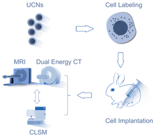
Monitoring implanted stem cells in bone regeneration and other cell therapies is of great importance to reveal the mechanism of tissue repair and to optimize clinical treatments. However, big challenge still remained in lacking an imaging nanoprobe. Herein, we designed surface modified upconversion nanoparticles (UCNs) with multimodal imaging capabilities of fluorescence, magnetic resonance imaging (MRI) and dual-energy computed tomography (CT). It was found that the UCNs can label stem cells in an efficient (over 200 pg/cell) and long-term (at least 14 days) manner, with almost no influence on the viability, cell cycle, apoptosis, and multilineage differentiation. Thus, clinical dual-energy CT and MRI were successfully applied to observe the migration of labeled cells on a bone-defect model of rabbit for at least 14 days. The results visualized the gathering of stem cells at the defect site of cortical bone, and the in vivo images were well-correlated with the in vitro fluorescence observation without extra staining. Therefore, a potentially translatable nanoprobe was developed for noninvasive and real-time tracking of cells, which may be meaningful for understanding the bone regeneration in clinic and shed light on the visualization of cells in other cell therapies.
中文翻译:

基于上转换纳米粒子的多峰纳米探针监测大动物骨缺损中植入的干细胞
监测植入的干细胞在骨再生和其他细胞疗法中的重要性对于揭示组织修复的机制和优化临床治疗非常重要。但是,仍然缺乏成像纳米探针仍然是一个巨大的挑战。在本文中,我们设计了具有荧光,磁共振成像(MRI)和双能计算机断层扫描(CT)的多峰成像功能的表面修饰的上转换纳米粒子(UCN)。发现UCN可以高效(超过200 pg /细胞)和长期(至少14天)的方式标记干细胞,而对存活率,细胞周期,细胞凋亡和多系分化几乎没有影响。因此,临床双能CT和MRI成功应用于至少14天的兔骨缺损模型上标记细胞的迁移。结果可视化了干细胞在皮质骨缺损部位的聚集,并且体内图像与体外荧光观察很好地相关,而没有额外的染色。因此,开发了一种潜在可翻译的纳米探针,用于细胞的非侵入性和实时跟踪,这对于理解临床中的骨再生以及为其他细胞疗法中的细胞可视化提供启示可能是有意义的。
更新日期:2018-01-23
中文翻译:
基于上转换纳米粒子的多峰纳米探针监测大动物骨缺损中植入的干细胞
监测植入的干细胞在骨再生和其他细胞疗法中的重要性对于揭示组织修复的机制和优化临床治疗非常重要。但是,仍然缺乏成像纳米探针仍然是一个巨大的挑战。在本文中,我们设计了具有荧光,磁共振成像(MRI)和双能计算机断层扫描(CT)的多峰成像功能的表面修饰的上转换纳米粒子(UCN)。发现UCN可以高效(超过200 pg /细胞)和长期(至少14天)的方式标记干细胞,而对存活率,细胞周期,细胞凋亡和多系分化几乎没有影响。因此,临床双能CT和MRI成功应用于至少14天的兔骨缺损模型上标记细胞的迁移。结果可视化了干细胞在皮质骨缺损部位的聚集,并且体内图像与体外荧光观察很好地相关,而没有额外的染色。因此,开发了一种潜在可翻译的纳米探针,用于细胞的非侵入性和实时跟踪,这对于理解临床中的骨再生以及为其他细胞疗法中的细胞可视化提供启示可能是有意义的。


 京公网安备 11010802027423号