Analytical and Bioanalytical Chemistry ( IF 3.8 ) Pub Date : 2017-11-28 , DOI: 10.1007/s00216-017-0739-2 Michiko Oya , Hiromi Suzuki , Andrea Roxanne J. Anas , Koichi Oishi , Kenji Ono , Shun Yamaguchi , Megumi Eguchi , Makoto Sawada
Mass spectrometry (MS) imaging is a useful tool for direct and simultaneous visualization of specific molecules. Liquid chromatography-tandem mass spectrometry (LC-MS/MS) is used to evaluate the abundance of molecules in tissues using sample homogenates. To date, however, LC-MS/MS has not been utilized as an imaging tool because spatial information is lost during sample preparation. Here we report a new approach for LC-MS/MS imaging using a thermal film-based laser microdissection (LMD) technique. To isolate tissue spots, our LMD system uses a 808-nm near infrared laser, the diameter of which can be freely changed from 2.7 to 500 μm; for imaging purposes in this study, the diameter was fixed at 40 μm, allowing acquisition of LC-MS/MS images at a 40-μm resolution. The isolated spots are arranged on a thermal film at 4.5-mm intervals, corresponding to the well spacing on a 384-well plate. Each tissue spot is handled on the film in such a manner as to maintain its spatial information, allowing it to be extracted separately in its individual well. Using analytical LC-MS/MS in combination with the spatial information of each sample, we can reconstruct LC-MS/MS images. With this imaging technique, we successfully obtained the distributions of pilocarpine, glutamate, γ-aminobutyric acid, acetylcholine, and choline in a cross-section of mouse hippocampus. The protocol we established in this study is applicable to revealing the neurochemistry of pilocarpine model of epilepsy. Our system has a wide range of uses in fields such as biology, pharmacology, pathology, and neuroscience.
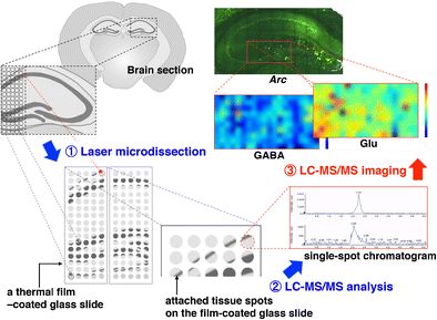
Schematic Indication of LMD-LC-MS/MS imaging.
中文翻译:
基于热膜激光显微切割的LC-MS / MS成像
质谱(MS)成像是直接和同时可视化特定分子的有用工具。液相色谱-串联质谱(LC-MS / MS)用于使用样品匀浆物评估组织中分子的丰度。但是,迄今为止,LC-MS / MS尚未被用作成像工具,因为在样品制备过程中会丢失空间信息。在这里,我们报告了一种使用基于热膜的激光显微切割(LMD)技术进行LC-MS / MS成像的新方法。为了隔离组织斑点,我们的LMD系统使用808 nm近红外激光,其直径可以自由地从2.7更改为500μm。为了本研究中的成像目的,直径固定为40μm,从而可以以40μm的分辨率采集LC-MS / MS图像。隔离的点以4.5毫米的间隔排列在热敏胶片上,对应于384孔板上的孔间距。以保持其空间信息的方式在胶片上处理每个组织点,从而使其分别在其单独的孔中被提取。使用分析型LC-MS / MS结合每个样品的空间信息,我们可以重建LC-MS / MS图像。通过这种成像技术,我们成功地获得了小鼠海马横截面上毛果芸香碱,谷氨酸,γ-氨基丁酸,乙酰胆碱和胆碱的分布。我们在这项研究中建立的协议适用于揭示毛果芸香碱癫痫模型的神经化学。我们的系统在生物学,药理学,病理学和神经科学等领域具有广泛的用途。以保持其空间信息的方式在胶片上处理每个组织点,从而使其分别在其单独的孔中被提取。使用分析型LC-MS / MS结合每个样品的空间信息,我们可以重建LC-MS / MS图像。通过这种成像技术,我们成功地获得了小鼠海马横截面上毛果芸香碱,谷氨酸,γ-氨基丁酸,乙酰胆碱和胆碱的分布。我们在这项研究中建立的协议适用于揭示毛果芸香碱癫痫模型的神经化学。我们的系统在生物学,药理学,病理学和神经科学等领域具有广泛的用途。以保持其空间信息的方式在胶片上处理每个组织点,从而使其分别在其单独的孔中被提取。使用分析型LC-MS / MS结合每个样品的空间信息,我们可以重建LC-MS / MS图像。通过这种成像技术,我们成功地获得了小鼠海马横截面上毛果芸香碱,谷氨酸,γ-氨基丁酸,乙酰胆碱和胆碱的分布。我们在这项研究中建立的协议适用于揭示毛果芸香碱癫痫模型的神经化学。我们的系统在生物学,药理学,病理学和神经科学等领域具有广泛的用途。使用分析型LC-MS / MS结合每个样品的空间信息,我们可以重建LC-MS / MS图像。通过这种成像技术,我们成功地获得了小鼠海马横截面上毛果芸香碱,谷氨酸,γ-氨基丁酸,乙酰胆碱和胆碱的分布。我们在这项研究中建立的协议适用于揭示毛果芸香碱癫痫模型的神经化学。我们的系统在生物学,药理学,病理学和神经科学等领域具有广泛的用途。使用分析型LC-MS / MS结合每个样品的空间信息,我们可以重建LC-MS / MS图像。通过这种成像技术,我们成功地获得了小鼠海马横截面上毛果芸香碱,谷氨酸,γ-氨基丁酸,乙酰胆碱和胆碱的分布。我们在这项研究中建立的协议适用于揭示毛果芸香碱癫痫模型的神经化学。我们的系统在生物学,药理学,病理学和神经科学等领域具有广泛的用途。我们在这项研究中建立的协议适用于揭示毛果芸香碱癫痫模型的神经化学。我们的系统在生物学,药理学,病理学和神经科学等领域具有广泛的用途。我们在这项研究中建立的协议适用于揭示毛果芸香碱癫痫模型的神经化学。我们的系统在生物学,药理学,病理学和神经科学等领域具有广泛的用途。
LMD-LC-MS / MS成像的示意图。


 京公网安备 11010802027423号