当前位置:
X-MOL 学术
›
Mol. Pharmaceutics
›
论文详情
Our official English website, www.x-mol.net, welcomes your
feedback! (Note: you will need to create a separate account there.)
Intratumoral Visualization of Oxaliplatin within a Liposomal Formulation Using X-ray Fluorescence Spectrometry
Molecular Pharmaceutics ( IF 4.5 ) Pub Date : 2018-01-16 00:00:00 , DOI: 10.1021/acs.molpharmaceut.7b00762 Hidenori Ando 1 , Amr S. Abu Lila 1, 2, 3 , Masao Tanaka 1 , Yusuke Doi 1 , Yasuko Terada 4 , Naoto Yagi 4 , Taro Shimizu 1 , Keiichiro Okuhira 1 , Yu Ishima 1 , Tatsuhiro Ishida 1
Molecular Pharmaceutics ( IF 4.5 ) Pub Date : 2018-01-16 00:00:00 , DOI: 10.1021/acs.molpharmaceut.7b00762 Hidenori Ando 1 , Amr S. Abu Lila 1, 2, 3 , Masao Tanaka 1 , Yusuke Doi 1 , Yasuko Terada 4 , Naoto Yagi 4 , Taro Shimizu 1 , Keiichiro Okuhira 1 , Yu Ishima 1 , Tatsuhiro Ishida 1
Affiliation

|
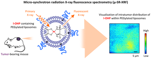
Microsynchrotron radiation X-ray fluorescence spectrometry (μ-SR-XRF) is an X-ray procedure that utilizes synchrotron radiation as an excitation source. μ-SR-XRF is a rapid, nondestructive technique that allows mapping and quantification of metals and biologically important elements in cell or tissue samples. Generally, the intratumor distribution of nanocarrier-based therapeutics is assessed by tracing the distribution of a labeled nanocarrier within tumor tissue, rather than by tracing the encapsulated drug. Instead of targeting the delivery vehicle, we employed μ-SR-XRF to visualize the intratumoral microdistribution of oxaliplatin (l-OHP) encapsulated within PEGylated liposomes. Tumor-bearing mice were intravenously injected with either l-OHP-containing PEGylated liposomes (l-OHP liposomes) or free l-OHP. The intratumor distribution of l-OHP within tumor sections was determined by detecting the fluorescence of platinum atoms, which are the main elemental components of l-OHP. The l-OHP in the liposomal formulation was localized near the tumor vessels and accumulated in tumors at concentrations greater than those seen with the free form, which is consistent with the results of our previous study that focused on fluorescent labeling of PEGylated liposomes. In addition, repeated administration of l-OHP liposomes substantially enhanced the tumor accumulation and/or intratumor distribution of a subsequent dose of l-OHP liposomes, presumably via improvements in tumor vascular permeability, which is also consistent with our previous results. In conclusion, μ-SR-XRF imaging efficiently and directly traced the intratumor distribution of the active pharmaceutical ingredient l-OHP encapsulated in liposomes within tumor tissue. μ-SR-XRF imaging could be a powerful means for estimating tissue distribution and even predicting the pharmacological effect of nanocarrier-based anticancer metal compounds.
中文翻译:

使用X射线荧光光谱法在脂质体制剂中奥沙利铂的瘤内可视化
微同步辐射X射线荧光光谱法(μ-SR-XRF)是一种利用同步辐射作为激发源的X射线程序。μ-SR-XRF是一种快速,无损的技术,可以对细胞或组织样品中的金属和生物学上重要的元素进行定位和定量。通常,通过追踪标记的纳米载体在肿瘤组织内的分布,而不是通过追踪包封的药物,来评估基于纳米载体的治疗剂的肿瘤内分布。代替靶向递送载体,我们采用μ-SR-XRF可视化封装在聚乙二醇化脂质体内的奥沙利铂(1-OHP)的肿瘤内微分布。给荷瘤小鼠静脉内注射含1-OHP的PEG化脂质体(1-OHP脂质体)或游离的1-OHP。通过检测铂原子的荧光来确定1-OHP在肿瘤切片内的肿瘤内分布,所述铂原子是1-OHP的主要元素组分。脂质体制剂中的l-OHP定位在肿瘤血管附近,并以比游离形式更大的浓度聚集在肿瘤中,这与我们先前专注于聚乙二醇化脂质体荧光标记的研究结果一致。另外,重复施用1-OHP脂质体大概可以通过改善肿瘤血管的通透性来增强随后剂量的1-OHP脂质体的肿瘤蓄积和/或肿瘤内分布,这也与我们先前的结果一致。综上所述,μ-SR-XRF成像可有效并直接追踪封装在肿瘤组织内脂质体内的活性药物成分1-OHP在肿瘤内的分布。μ-SR-XRF成像可能是估计组织分布甚至预测基于纳米载体的抗癌金属化合物的药理作用的有力手段。
更新日期:2018-01-16
中文翻译:

使用X射线荧光光谱法在脂质体制剂中奥沙利铂的瘤内可视化
微同步辐射X射线荧光光谱法(μ-SR-XRF)是一种利用同步辐射作为激发源的X射线程序。μ-SR-XRF是一种快速,无损的技术,可以对细胞或组织样品中的金属和生物学上重要的元素进行定位和定量。通常,通过追踪标记的纳米载体在肿瘤组织内的分布,而不是通过追踪包封的药物,来评估基于纳米载体的治疗剂的肿瘤内分布。代替靶向递送载体,我们采用μ-SR-XRF可视化封装在聚乙二醇化脂质体内的奥沙利铂(1-OHP)的肿瘤内微分布。给荷瘤小鼠静脉内注射含1-OHP的PEG化脂质体(1-OHP脂质体)或游离的1-OHP。通过检测铂原子的荧光来确定1-OHP在肿瘤切片内的肿瘤内分布,所述铂原子是1-OHP的主要元素组分。脂质体制剂中的l-OHP定位在肿瘤血管附近,并以比游离形式更大的浓度聚集在肿瘤中,这与我们先前专注于聚乙二醇化脂质体荧光标记的研究结果一致。另外,重复施用1-OHP脂质体大概可以通过改善肿瘤血管的通透性来增强随后剂量的1-OHP脂质体的肿瘤蓄积和/或肿瘤内分布,这也与我们先前的结果一致。综上所述,μ-SR-XRF成像可有效并直接追踪封装在肿瘤组织内脂质体内的活性药物成分1-OHP在肿瘤内的分布。μ-SR-XRF成像可能是估计组织分布甚至预测基于纳米载体的抗癌金属化合物的药理作用的有力手段。


 京公网安备 11010802027423号