Talanta ( IF 5.6 ) Pub Date : 2017-12-27 , DOI: 10.1016/j.talanta.2017.12.066 Rongxin Fu , Qi Li , Ruliang Wang , Ning Xue , Xue Lin , Ya Su , Kai Jiang , Xiangyu Jin , Rongzan Lin , Wupeng Gan , Ying Lu , Guoliang Huang
|
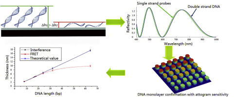
Interferometric imaging biosensors are powerful and convenient tools for confirming the existence of DNA monolayer films on silicon microarray platforms. However, their accuracy and sensitivity need further improvement because DNA molecules contribute to an inconspicuous interferometric signal both in thickness and size. Such weaknesses result in poor performance of these biosensors for low DNA content analyses and point mutation tests. In this paper, an interferometric imaging biosensor with weighted spectrum analysis is presented to confirm DNA monolayer films. The interferometric signal of DNA molecules can be extracted and then quantitative detection results for DNA microarrays can be reconstructed. With the proposed strategy, the relative error of thickness detection was reduced from 88.94% to merely 4.15%. The mass sensitivity per unit area of the proposed biosensor reached 20 attograms (ag). Therefore, the sample consumption per unit area of the target DNA content was only 62.5 zeptomoles (zm), with the volume of 0.25 picolitres (pL). Compared with the fluorescence resonance energy transfer (FRET), the measurement veracity of the interferometric imaging biosensor with weighted spectrum analysis is free to the changes in spotting concentration and DNA length. The detection range was more than 1 µm. Moreover, single nucleotide mismatch could be pointed out combined with specific DNA ligation. A mutation experiment for lung cancer detection proved the high selectivity and accurate analysis capability of the presented biosensor.
中文翻译:
干涉成像生物传感器,使用加权光谱分析来确认具有ATTAGRAPH敏感性的DNA单层膜
干涉成像生物传感器是功能强大且方便的工具,可用于确认硅微阵列平台上DNA单层膜的存在。但是,由于DNA分子在厚度和尺寸上都会引起不起眼的干涉信号,因此它们的准确性和灵敏性需要进一步提高。这些弱点导致这些生物传感器在低DNA含量分析和点突变测试中的性能较差。在本文中,提出了一种具有加权光谱分析的干涉成像生物传感器,以确认DNA单层膜。可以提取DNA分子的干涉信号,然后可以重建DNA微阵列的定量检测结果。通过提出的策略,厚度检测的相对误差从88.94%降低到仅4.15%。所提出的生物传感器每单位面积的质量灵敏度达到20 ATTG(ag)。因此,目标DNA含量每单位面积的样品消耗量仅为62.5 zeptomoles(zm),体积为0.25皮升(pL)。与荧光共振能量转移(FRET)相比,采用加权光谱分析的干涉成像生物传感器的测量准确性不受斑点浓度和DNA长度变化的影响。检测范围大于1μm。而且,可以指出单核苷酸错配与特异性DNA连接相结合。用于肺癌检测的突变实验证明了该生物传感器的高选择性和准确的分析能力。单位面积目标DNA含量的样品消耗量仅为62.5泽摩尔(zm),体积为0.25皮升(pL)。与荧光共振能量转移(FRET)相比,采用加权光谱分析的干涉成像生物传感器的测量准确性不受斑点浓度和DNA长度变化的影响。检测范围大于1μm。而且,可以指出单核苷酸错配与特异性DNA连接相结合。用于肺癌检测的突变实验证明了该生物传感器的高选择性和准确的分析能力。单位面积目标DNA含量的样品消耗量仅为62.5泽摩尔(zm),体积为0.25皮升(pL)。与荧光共振能量转移(FRET)相比,采用加权光谱分析的干涉成像生物传感器的测量准确性不受斑点浓度和DNA长度变化的影响。检测范围大于1μm。而且,可以指出单核苷酸错配与特异性DNA连接相结合。用于肺癌检测的突变实验证明了该生物传感器的高选择性和准确的分析能力。干涉光谱成像生物传感器的加权光谱分析的测量准确性不受斑点浓度和DNA长度变化的影响。检测范围大于1μm。而且,可以指出单核苷酸错配与特异性DNA连接相结合。用于肺癌检测的突变实验证明了该生物传感器的高选择性和准确的分析能力。干涉光谱成像生物传感器的加权光谱分析的测量准确性不受斑点浓度和DNA长度变化的影响。检测范围大于1μm。而且,可以指出单核苷酸错配与特异性DNA连接相结合。用于肺癌检测的突变实验证明了该生物传感器的高选择性和准确的分析能力。


 京公网安备 11010802027423号