Acta Biomaterialia ( IF 9.7 ) Pub Date : 2017-12-27 , DOI: 10.1016/j.actbio.2017.12.031 Victor Balashov , Anton Efimov , Olga Agapova , Alexander Pogorelov , Igor Agapov , Konstantin Agladze
|
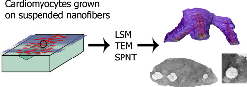
Building functional and robust scaffolds for engineered biological tissue requires a nanoscale mechanistic understanding of how cells use the scaffold for their growth and development. A vast majority of the scaffolds used for cardiac tissue engineering are based on polymer materials, the matrices of nanofibers. Attempts to load the polymer fibers of the scaffold with additional sophisticated features, such as electrical conductivity and controlled release of the growth factors or other biologically active molecules, as well as trying to match the mechanical features of the scaffold to those of the extracellular matrix, cannot be efficient without a detailed knowledge of how the cells are attached and strategically positioned with respect to the scaffold nanofibers at micro and nanolevel. Studying single cell – single fiber interactions with the aid of confocal laser scanning microscopy (CLSM), scanning probe nanotomography (SPNT), and transmission electron microscopy (TEM), we found that cardiac cells actively interact with substrate nanofibers, but in different ways. While cardiomyocytes often create a remarkable “sheath” structure, enveloping fiber and, thus, substantially increasing contact zone, fibroblasts interact with nanofibers in the locations of focal adhesion clusters mainly without wrapping the fiber.
Statements of Significance
We found that cardiomyocytes grown on electrospun polymer nanofibers often create a striking “sheath” structure, enveloping fiber with the formation of a very narrow (∼22 nm) membrane gap leading from the fiber to the extracellular space. This wrapping makes the entire fiber surface available for cell attachment. This finding gives a new prospective view on how scaffold nanofibers may interact with growing cells. It may play a significant role in effective design of novel nanofiber scaffolds for tissue engineering concerning mechanical and electrical properties of scaffolds as well as controlled drug release from “smart” biomaterials.
中文翻译:
聚合物支架纳米纤维上心肌细胞的高分辨率3D显微镜研究显示异常鞘结构的形成
为工程化的生物组织构建功能强大且功能强大的支架,需要对细胞如何使用支架进行生长和发育的纳米尺度的机械理解。用于心脏组织工程的绝大多数支架都基于聚合物材料,即纳米纤维的基质。尝试为支架的聚合物纤维加载其他复杂功能,例如电导率和生长因子或其他生物活性分子的受控释放,以及尝试使支架的机械特征与细胞外基质的机械特征匹配,没有详细了解细胞如何相对于支架纳米纤维在微米和纳米水平上附着和策略性定位的详细信息,就无法实现高效。在共聚焦激光扫描显微镜(CLSM),扫描探针纳米断层扫描(SPNT)和透射电子显微镜(TEM)的帮助下研究单细胞-单纤维相互作用,我们发现心脏细胞与底物纳米纤维活跃地相互作用,但是方式不同。尽管心肌细胞通常会形成一个显着的“护套”结构,包裹着纤维,从而大大增加了接触区域,但成纤维细胞在粘着斑簇的位置与纳米纤维相互作用,而基本上没有包裹纤维。
重要声明
我们发现,在静电纺丝聚合物纳米纤维上生长的心肌细胞通常会形成引人注目的“鞘”结构,将纤维包裹起来,形成从纤维到细胞外空间的非常狭窄的膜间隙(〜22 nm)。这种包裹使整个纤维表面可用于细胞附着。这一发现为支架纳米纤维如何与生长中的细胞相互作用提供了新的前瞻性观点。它在有效设计用于组织工程的新型纳米纤维支架方面可能发挥重要作用,涉及支架的机械和电气特性以及从“智能”生物材料中控制药物的释放。


 京公网安备 11010802027423号