Analytical and Bioanalytical Chemistry ( IF 4.3 ) Pub Date : 2017-12-26 , DOI: 10.1007/s00216-017-0837-1 Jia Jiang , Sizhu Tian , Kun Wang , Yang Wang , Shuang Zang , Aimin Yu , Ziwei Zhang
With the help of iron oxide nanoparticles, electron spin resonance spectroscopy (ESR) was applied to immunoassay. Iron oxide nanoparticles were used as the ESR probe in order to achieve an amplification of the signal resulting from the large amount of Fe3+ ion enclosed in each nanoparticle. Rabbit IgG was used as antigen to test this method. Polyclonal antibody of rabbit IgG was used as antibody to detect the antigen. Iron oxide nanoparticle with a diameter of either 10 or 30 nm was labeled to the antibody, and Fe3+ in the nanoparticle was probed for ESR signal. The sepharose beads were used as solid phase to which rabbit IgG was conjugated. The nanoparticle-labeled antibody was first added in the sample containing antigen, and the antigen-conjugated sepharose beads were then added into the sample. The nanoparticle-labeled antibody bound to the antigen on sepharose beads was separated from the sample by centrifugation and measured. We found that the detection ranges of the antigen obtained with nanoparticles of different sizes were different because the amount of antibody on nanoparticles of 10 nm was about one order of magnitude higher than that on nanoparticles of 30 nm. When 10 nm nanoparticle was used as probe, the upper limit of detection was 40.00 μg mL−1, and the analytical sensitivity was 1.81 μg mL−1. When 30 nm nanoparticle was used, the upper limit of detection was 3.00 μg mL−1, and the sensitivity was 0.014 and 0.13 μg mL−1 depending on the ratio of nanoparticle to antibody.
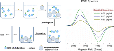
Schematic diagram of procedure and ESR spectra
中文翻译:
电子自旋共振光谱用于氧化铁纳米粒子作为探针的免疫测定
借助氧化铁纳米颗粒,将电子自旋共振光谱(ESR)应用于免疫测定。氧化铁纳米粒子被用作ESR探针,以实现放大每个纳米粒子中包裹的大量Fe 3+离子产生的信号的功能。兔IgG用作抗原来测试该方法。兔IgG多克隆抗体被用作检测抗原的抗体。直径为10或30 nm的氧化铁纳米颗粒被标记为抗体,并且Fe 3+探测纳米粒子中的ESR信号。琼脂糖凝胶珠用作与兔IgG缀合的固相。首先将纳米颗粒标记的抗体添加到含有抗原的样品中,然后将抗原结合的琼脂糖珠添加到样品中。通过离心将与琼脂糖珠上的抗原结合的纳米颗粒标记的抗体与样品分离并进行测量。我们发现,用不同大小的纳米颗粒获得的抗原的检测范围是不同的,因为10 nm纳米颗粒上的抗体量比30 nm纳米颗粒上的抗体量高约一个数量级。当使用10 nm纳米颗粒作为探针时,检测上限为40.00μgmL -1,分析灵敏度为1.81μgmL-1。当使用30nm纳米颗粒时,取决于纳米颗粒与抗体的比例,检测的上限为3.00μgmL -1,并且灵敏度为0.014和0.13μgmL -1。
步骤和ESR谱图


 京公网安备 11010802027423号