Our official English website, www.x-mol.net, welcomes your
feedback! (Note: you will need to create a separate account there.)
Shape-Dependent Dissolution and Cellular Uptake of Silver Nanoparticles
Langmuir ( IF 3.7 ) Pub Date : 2018-01-16 00:00:00 , DOI: 10.1021/acs.langmuir.7b03126 Christina Graf 1 , Daniel Nordmeyer 1 , Christina Sengstock 2 , Sebastian Ahlberg 3 , Jörg Diendorf 4 , Jörg Raabe 5 , Matthias Epple 4 , Manfred Köller 2 , Jürgen Lademann 3 , Annika Vogt 3 , Fiorenza Rancan 3 , Eckart Rühl 1
Langmuir ( IF 3.7 ) Pub Date : 2018-01-16 00:00:00 , DOI: 10.1021/acs.langmuir.7b03126 Christina Graf 1 , Daniel Nordmeyer 1 , Christina Sengstock 2 , Sebastian Ahlberg 3 , Jörg Diendorf 4 , Jörg Raabe 5 , Matthias Epple 4 , Manfred Köller 2 , Jürgen Lademann 3 , Annika Vogt 3 , Fiorenza Rancan 3 , Eckart Rühl 1
Affiliation

|
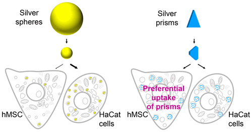
The cellular uptake and dissolution of trigonal silver nanoprisms (edge length 42 ± 15 nm, thickness 8 ± 1 nm) and mostly spherical silver nanoparticles (diameter 70 ± 25 nm) in human mesenchymal stem cells (hMSC’s) and human keratinocytes (HaCaT cells) were investigated. Both particles are stabilized by polyvinylpyrrolidone (PVP), with the prisms additionally stabilized by citrate. The nanoprisms dissolved slightly in pure water but strongly in isotonic saline or at pH 4, corresponding to the lowest limit for the pH during cellular uptake. The tips of the prisms became rounded within minutes due to their high surface energy. Afterward, the dissolution process slowed down due to the presence of both PVP stabilizing Ag{100} sites and citrate blocking Ag{111} sites. On the contrary, nanospheres, solely stabilized by PVP, dissolved within 24 h. These results correlate with the finding that particles in both cell types have lost >90% of their volume within 24 h. hMSC’s took up significantly more Ag from nanoprisms than from nanospheres, whereas HaCaT cells showed no preference for one particle shape. This can be rationalized by the large cellular interaction area of the plateletlike nanoprisms and the bending stiffness of the cell membranes. hMSC’s have a highly flexible cell membrane, resulting in an increased uptake of plateletlike particles. HaCaT cells have a membrane with a 3 orders of magnitude higher Young’s modulus than for hMSC. Hence, the energy gain due to the larger interaction area of the nanoprisms is compensated for by the higher energy needed for cell membrane deformation compared to that for spheres, leading to no shape preference.
中文翻译:

银纳米颗粒的形状依赖性溶解和细胞吸收
在人间充质干细胞(hMSC's)和人角质形成细胞(HaCaT细胞)中,三角形的银纳米棱镜(边缘长度42±15 nm,厚度8±1 nm)以及大部分球形银纳米颗粒(直径70±25 nm)的细胞吸收和溶解被调查了。两种颗粒均通过聚乙烯吡咯烷酮(PVP)稳定,而棱镜另外还通过柠檬酸盐稳定。纳米棱镜在纯水中略微溶解,但在等渗盐水中或在pH 4时强烈溶解,这对应于细胞摄取过程中pH的最低极限。棱镜的尖端由于其较高的表面能而在数分钟内就变圆了。之后,由于同时存在PVP稳定Ag {100}位点和柠檬酸盐阻断Ag {111}位点,溶解过程变慢了。相反,仅由PVP稳定的纳米球在24小时内溶解。这些结果与以下发现相关:两种细胞类型中的颗粒在24小时内损失了> 90%的体积。hMSC从纳米棱镜中吸收的银要比从纳米球吸收的银多得多,而HaCaT细胞对一种粒子的形状没有偏爱。这可以通过血小板状纳米棱镜的大细胞相互作用区域和细胞膜的弯曲刚度来合理化。hMSC具有高度柔性的细胞膜,导致血小板样颗粒的摄取增加。HaCaT细胞的膜的杨氏模量比hMSC高3个数量级。因此,与球形相比,由于纳米棱柱的较大相互作用区域而产生的能量增益被细胞膜变形所需的较高能量所补偿,从而导致没有形状偏爱。
更新日期:2018-01-16
中文翻译:

银纳米颗粒的形状依赖性溶解和细胞吸收
在人间充质干细胞(hMSC's)和人角质形成细胞(HaCaT细胞)中,三角形的银纳米棱镜(边缘长度42±15 nm,厚度8±1 nm)以及大部分球形银纳米颗粒(直径70±25 nm)的细胞吸收和溶解被调查了。两种颗粒均通过聚乙烯吡咯烷酮(PVP)稳定,而棱镜另外还通过柠檬酸盐稳定。纳米棱镜在纯水中略微溶解,但在等渗盐水中或在pH 4时强烈溶解,这对应于细胞摄取过程中pH的最低极限。棱镜的尖端由于其较高的表面能而在数分钟内就变圆了。之后,由于同时存在PVP稳定Ag {100}位点和柠檬酸盐阻断Ag {111}位点,溶解过程变慢了。相反,仅由PVP稳定的纳米球在24小时内溶解。这些结果与以下发现相关:两种细胞类型中的颗粒在24小时内损失了> 90%的体积。hMSC从纳米棱镜中吸收的银要比从纳米球吸收的银多得多,而HaCaT细胞对一种粒子的形状没有偏爱。这可以通过血小板状纳米棱镜的大细胞相互作用区域和细胞膜的弯曲刚度来合理化。hMSC具有高度柔性的细胞膜,导致血小板样颗粒的摄取增加。HaCaT细胞的膜的杨氏模量比hMSC高3个数量级。因此,与球形相比,由于纳米棱柱的较大相互作用区域而产生的能量增益被细胞膜变形所需的较高能量所补偿,从而导致没有形状偏爱。


 京公网安备 11010802027423号