Acta Biomaterialia ( IF 9.4 ) Pub Date : 2017-12-21 , DOI: 10.1016/j.actbio.2017.12.022 Jin-long Sun , Kai Jiao , Qun Song , Chu-fan Ma , Chao Ma , Franklin R. Tay , Li-na Niu , Ji-hua Chen
|
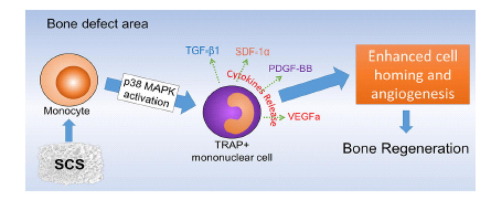
Intrafibrillar silicified collagen scaffold (SCS) is a promising biomaterial for bone regeneration because it promotes cell homing and angiogenesis in bone defects via monocyte modulation. In the present study, a rat femoral defect model was used to examine the contribution of monocyte signaling pathways to SCS modulation. Activation of the monocyte p38 signaling pathway by SCS resulted in monocyte differentiation into TRAP-positive mononuclear cells. These cells demonstrated increased secretion of SDF-1α, VEGFa and PDGF-BB, which, in turn, promoted homing of bone marrow stromal cells (BMSCs) and endothelial progenitor cells (EPCs), as well as local vascularization. Monocyte differentiation and secretion were blocked after inhibition of the p38 pathway, which resulted in reduction in cell homing and angiogenesis. Taken together, these novel findings indicate that the p38 signaling pathway is crucial in SCS-modulated monocyte differentiation and secretion, which has a direct impact on SCS-induced bone regeneration.
Statement of significance
Intrafibrillar silicified collagen scaffold (SCS) is a promising biomaterial for bone regeneration. The present work demonstrates that SCS possesses favorable bone regeneration potential in a rat femoral defect model. The degrading scaffold modulates monocyte differentiation and release of certain cytokines to recruit MSCs and EPCs, as well as enhances local vascularization by activating the p38 MAPK signaling pathway. These findings indicate that SCS contributes to bone defect regeneration by stimulating host cell homing and promoting local angiogenesis and osteogenesis without the need for loading cytokines or xenogenous stem cells.
中文翻译:
纤维内硅化胶原蛋白支架通过激活单核细胞p38信号通路促进原位骨再生
纤维内硅化胶原蛋白支架(SCS)是一种有前途的骨再生生物材料,因为它通过单核细胞调节促进骨缺损中的细胞归巢和血管生成。在本研究中,使用大鼠股骨缺损模型检查单核细胞信号通路对SCS调节的贡献。SCS对单核细胞p38信号通路的激活导致单核细胞分化为TRAP阳性单核细胞。这些细胞显示出SDF-1α,VEGFa和PDGF-BB的分泌增加,进而促进了骨髓基质细胞(BMSC)和内皮祖细胞(EPC)的归巢,以及局部血管形成。抑制p38途径后,单核细胞的分化和分泌被阻断,这导致细胞归巢和血管生成减少。在一起
重要声明
纤维内硅化胶原蛋白支架(SCS)是一种有前途的骨再生生物材料。目前的工作表明,SCS在大鼠股骨缺损模型中具有良好的骨再生潜力。降解支架调节单核细胞的分化和某些细胞因子的释放以募集MSC和EPC,并通过激活p38 MAPK信号通路增强局部血管形成。这些发现表明,SCS通过刺激宿主细胞归巢并促进局部血管生成和成骨而有助于骨缺损的再生,而无需加载细胞因子或异种干细胞。


 京公网安备 11010802027423号