Acta Biomaterialia ( IF 9.4 ) Pub Date : 2017-12-19 , DOI: 10.1016/j.actbio.2017.12.018 Jiang Jiang 1 , Shixuan Chen 1 , Hongjun Wang 1 , Mark A Carlson 2 , Adrian F Gombart 3 , Jingwei Xie 1
|
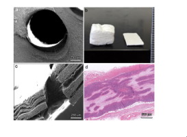
Traditional electrospun nanofiber membranes were incapable of promoting cellular infiltration due to its intrinsic property (e.g., dense structure and small pore size) limiting their use in tissue regeneration. Herein, we report a simple and novel approach for expanding traditional nanofiber membranes from two-dimensional to three-dimensional (3D) with controlled thickness and porosity via depressurization of subcritical CO2 fluid. The expanded 3D nanofiber scaffolds formed layered structures and simultaneously maintained the aligned nanotopographic cues. The 3D scaffolds also retained the fluorescent intensity of encapsulated coumarin 6 and the antibacterial activity of encapsulated antimicrobial peptide LL-37. In addition, the expanded 3D nanofiber scaffolds with arrayed holes can significantly promote cellular infiltration and neotissue formation after subcutaneous implantation compared to traditional nanofiber membranes. Such scaffolds also significantly increased the blood vessel formation and the ratio of M2/M1 macrophages after subcutaneous implantation for 2 and 4 weeks compared to traditional nanofiber membranes. Together, the presented method holds great potential in the fabrication of functional 3D nanofiber scaffolds for various applications including engineering 3D in vitro tissue models, antimicrobial wound dressing, and repairing/regenerating tissues in vivo.
Statement of Significance
Electrospun nanofibers have been widely used in regenerative medicine due to its biomimicry property. However, most of studies are limited to the use of 2D electrospun nanofiber membranes. To the best of our knowledge, this article is the first instance of the transformation of traditional electrospun nanofiber membranes from 2D to 3D via depressurization of subcritical CO2 fluid. This method eliminates many issues associated with previous approaches such as necessitating the use of aqueous solutions and chemical reactions, multiple-step process, loss of the activity of encapsulated biological molecules, and unable to expand electrospun nanofiber mats made of hydrophilic polymers. Results indicate that these CO2 expanded nanofiber scaffolds can maintain the activity of encapsulated biological molecules. Further, the CO2 expanded nanofiber scaffolds with arrayed holes can greatly promote cellular infiltration, neovascularization, and positive host response after subcutaneous implantation in rats. The current work is the first study elucidating such a simple and novel strategy for fabrication of 3D nanofiber scaffolds.
中文翻译:
CO2 膨胀纳米纤维支架保持封装生物活性材料的活性并促进细胞浸润和积极的宿主反应
传统的电纺纳米纤维膜由于其固有特性(例如致密结构和小孔径)而无法促进细胞浸润,限制了其在组织再生中的应用。在此,我们报告了一种简单而新颖的方法,通过亚临界CO 2流体减压,将传统纳米纤维膜从二维扩展到三维(3D),并控制厚度和孔隙率。扩展的 3D 纳米纤维支架形成分层结构,同时保持对齐的纳米形貌线索。 3D支架还保留了封装的香豆素6的荧光强度和封装的抗菌肽LL-37的抗菌活性。此外,与传统纳米纤维膜相比,具有阵列孔的扩展3D纳米纤维支架可以显着促进皮下植入后的细胞浸润和新组织形成。与传统的纳米纤维膜相比,这种支架在皮下植入2周和4周后还显着增加了血管形成和M2/M1巨噬细胞的比例。总之,所提出的方法在制造功能性 3D 纳米纤维支架方面具有巨大的潜力,可用于各种应用,包括工程 3D体外组织模型、抗菌伤口敷料和体内组织修复/再生。
重要性声明
静电纺纳米纤维由于其仿生特性而被广泛应用于再生医学领域。然而,大多数研究仅限于二维电纺纳米纤维膜的使用。据我们所知,本文是通过亚临界CO 2流体减压将传统静电纺纳米纤维膜从2D转变为3D的第一个实例。这种方法消除了与以前的方法相关的许多问题,例如需要使用水溶液和化学反应、多步骤过程、封装生物分子活性的损失以及无法扩展由亲水聚合物制成的电纺纳米纤维垫。结果表明,这些CO 2膨胀的纳米纤维支架可以保持封装的生物分子的活性。此外,具有阵列孔的CO 2膨胀纳米纤维支架在皮下植入大鼠体内后可以极大地促进细胞浸润、新生血管形成和积极的宿主反应。目前的工作是第一项阐明这种简单而新颖的 3D 纳米纤维支架制造策略的研究。


 京公网安备 11010802027423号