Journal of the Mechanical Behavior of Biomedical Materials ( IF 3.3 ) Pub Date : 2017-12-09 , DOI: 10.1016/j.jmbbm.2017.11.045 J.L. Schmidt , D.J. Tweten , A.A. Badachhape , A.J. Reiter , R.J. Okamoto , J.R. Garbow , P.V. Bayly
|
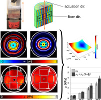
The mechanical properties of brain tissue, particularly those of white matter (WM), need to be characterized accurately for use in finite element (FE) models of brain biomechanics and traumatic brain injury (TBI). Magnetic resonance elastography (MRE) is a powerful tool for non-invasive estimation of the mechanical properties of soft tissues. While several studies involving direct mechanical tests of brain tissue have shown mechanical anisotropy, most MRE studies of brain tissue assume an isotropic model. In this study, an incompressible transversely isotropic (TI) material model parameterized by minimum shear modulus (), shear anisotropy parameter (), and tensile anisotropy parameter () is applied to analyze MRE measurements of ex vivo porcine white matter (WM) brain tissue. To characterize shear anisotropy, “slow” (pure transverse) shear waves were propagated at 100, 200 and 300 Hz through sections of ex vivo brain tissue including both WM and gray matter (GM). Shear waves were found to propagate with elliptical fronts, consistent with TI material behavior. Shear wave fields were also analyzed within regions of interest (ROI) to find local shear wavelengths parallel and perpendicular to fiber orientation. FE simulations of a TI material with a range of plausible shear modulus () and shear anisotropy parameters () were run and the results were analyzed in the same fashion as the experimental case. Parameters of the FE simulations which most closely matched each experiment were taken to represent the mechanical properties of that particular sample. Using this approach, WM in the ex vivo porcine brain was found to be mildly anisotropic in shear with estimates of minimum shear modulus (actuation frequencies listed in parenthesis): 1.04 0.12 kPa (at 100 Hz), 1.94 0.29 kPa (at 200 Hz), and 2.88 0.34 kPa (at 300 Hz) and corresponding shear anisotropy factors of 0.27 0.09 (at 100 Hz), 0.29 0.14 (at 200 Hz) and 0.34 0.13 (at 300 Hz). Future MRE studies will focus on tensile anisotropy, which will require both slow and fast shear waves for accurate estimation.
中文翻译:
磁共振弹性成像法体外测定猪脑白质中的各向异性力学性能
需要准确表征脑组织的机械性能,尤其是白质(WM)的机械性能,以用于脑生物力学和脑外伤(TBI)的有限元(FE)模型。磁共振弹性成像(MRE)是用于非侵入性评估软组织机械特性的强大工具。尽管涉及脑组织直接机械测试的多项研究表明机械各向异性,但大多数脑组织的MRE研究均采用各向同性模型。在这项研究中,通过最小剪切模量(),剪切各向异性参数()和拉伸各向异性参数()用于分析离体猪白质(WM)脑组织的MRE测量。为了表征剪切各向异性,“慢”(纯横向)剪切波以100、200和300 Hz的频率通过包括WM和灰质(GM)在内的离体脑组织切片传播。发现剪切波以椭圆形前沿传播,这与TI的材料行为一致。还对感兴趣区域(ROI)内的剪切波场进行了分析,以发现平行于和垂直于纤维取向的局部剪切波长。TI材料在一定范围内的合理剪切模量的有限元模拟()和剪切各向异性参数(),并以与实验案例相同的方式分析结果。采用与每个实验最匹配的有限元模拟参数,以表示该特定样品的机械性能。使用这种方法,发现离体猪脑中的WM在剪切中具有中等各向异性,并具有最小剪切模量(括号中列出的驱动频率)的估计值: 1.04 0.12 kPa(在100 Hz时), 1.94 0.29 kPa(在200 Hz时),和 2.88 0.34 kPa(在300 Hz时)和相应的剪切各向异性因子为 0.27 0.09(在100 Hz时), 0.29 0.14(在200 Hz时)和 0.34 0.13(在300 Hz时)。未来的MRE研究将集中于拉伸各向异性,这将需要慢速剪切波和快速剪切波来进行准确估计。


 京公网安备 11010802027423号