Acta Biomaterialia ( IF 9.7 ) Pub Date : 2017-12-12 , DOI: 10.1016/j.actbio.2017.12.008 Y. Li , J. Zhou , P. Pavanram , M.A. Leeflang , L.I. Fockaert , B. Pouran , N. Tümer , K.-U. Schröder , J.M.C. Mol , H. Weinans , H. Jahr , A.A. Zadpoor
|
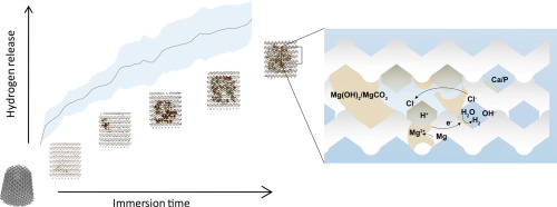
An ideal bone substituting material should be bone-mimicking in terms of mechanical properties, present a precisely controlled and fully interconnected porous structure, and degrade in the human body to allow for full regeneration of large bony defects. However, simultaneously satisfying all these three requirements has so far been highly challenging. Here we present topologically ordered porous magnesium (WE43) scaffolds based on the diamond unit cell that were fabricated by selective laser melting (SLM) and satisfy all the requirements. We studied the in vitro biodegradation behavior (up to 4 weeks), mechanical properties and biocompatibility of the developed scaffolds. The mechanical properties of the AM porous WE43 (E = 700-800 MPa) scaffolds were found to fall into the range of the values reported for trabecular bone even after 4 weeks of biodegradation. Scanning electron microscopy (SEM), Fourier transform infrared spectroscopy (FTIR), electrochemical tests and µCT revealed a unique biodegradation mechanism that started with uniform corrosion, followed by localized corrosion, particularly in the center of the scaffolds. Biocompatibility tests performed up to 72 h showed level 0 cytotoxicity (according to ISO 10993-5 and -12), except for one time point (i.e., 24 h). Intimate contact between cells (MG-63) and the scaffolds was also observed in SEM images. The study shows for the first time that AM of porous Mg may provide distinct possibilities to adjust biodegradation profile through topological design and open up unprecedented opportunities to develop multifunctional bone substituting materials that mimic bone properties and enable full regeneration of critical-size load-bearing bony defects.
Statement of significance
The ideal biomaterials for bone tissue regeneration should be bone-mimicking in terms of mechanical properties, present a fully interconnected porous structure, and exhibit a specific biodegradation behavior to enable full regeneration of bony defects. Recent advances in additive manufacturing have resulted in biomaterials that satisfy the first two requirements but simultaneously satisfying the third requirement has proven challenging so far. Here we present additively manufactured porous magnesium structures that have the potential to satisfy all above-mentioned requirements. Even after 4 weeks of biodegradation, the mechanical properties of the porous structures were found to be within those reported for native bone. Moreover, our comprehensive electrochemical, mechanical, topological, and biological study revealed a unique biodegradation behavior and the limited cytotoxicity of the developed biomaterials.
中文翻译:
增材制造的可生物降解的多孔镁
理想的骨替代材料应在机械性能方面模仿骨,呈现精确控制和完全互连的多孔结构,并在人体中降解以允许大骨缺损的完全再生。然而,到目前为止,同时满足所有这三个要求一直是极富挑战性的。在这里,我们介绍了基于金刚石晶胞的拓扑排序的多孔镁(WE43)支架,这些支架是通过选择性激光熔化(SLM)制成的,并且满足所有要求。我们研究了发达支架的体外生物降解行为(长达4周),机械性能和生物相容性。AM多孔WE43(E的力学性能。在经过4周的生物降解后,发现支架(= 700-800 MPa)落入了小梁骨报道的值范围内。扫描电子显微镜(SEM),傅立叶变换红外光谱(FTIR),电化学测试和µCT揭示了独特的生物降解机理,该机理始于均匀腐蚀,然后是局部腐蚀,特别是在支架的中央。进行长达72小时的生物相容性测试,除一个时间点(即24小时)外,细胞毒性均为0级(根据ISO 10993-5和-12)。在SEM图像中也观察到细胞(MG-63)与支架之间的紧密接触。
重要声明
用于骨组织再生的理想生物材料应在机械性能方面模仿骨,呈现出完全相互连接的多孔结构,并表现出特定的生物降解行为以实现充分的生物降解。骨缺损的再生。增材制造的最新进展已导致满足前两个要求但同时满足第三个要求的生物材料具有挑战性。在这里,我们介绍了可以满足所有上述要求的增材制造的多孔镁结构。即使经过4周的生物降解,多孔结构的机械性能仍被发现在天然骨的力学性能之内。此外,我们全面的电化学,机械,拓扑和生物学研究表明,这种独特的生物降解行为和已开发生物材料的有限细胞毒性。


 京公网安备 11010802027423号