Nano Research ( IF 9.9 ) Pub Date : 2018-08-02 , DOI: 10.1007/s12274-017-1934-3 Gu Cheng , Jiajia Chen , Qun Wang , Xuewen Yang , Yuet Cheng , Zhi Li , Hu Tu , Hongbing Deng , Zubing Li
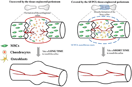
Various composite materials are now used as artificial tissue substitutes, and are defining new frontiers in tissue engineering. In the present study, composite membranes based on silk fibroin (SF) were fabricated to form a synthetic periosteum. The fabricated membranes were physicochemically characterized by their morphology, porosity, biocompatibility, biodegradability, chemical structure, and mechanical properties. Following the addition of polycaprolactone (PCL) to the silk fibers, there was a 3–5-fold increase in the elongation at break compared with the pure silk membranes, and surface wettability was retained. The degradation time of the SF within the membranes was also prolonged by adding PCL. Compared with pure PCL membranes or plastic culture plates, the SF-based membranes significantly enhanced the cellular viability and osteogenic differentiation capability of MC3T3-E1 cells. Higher expression levels of osteogenic differentiation markers (runt-related transcription factor 2 (RUNX2), alkaline phosphatase (ALP), and osteopontin (OP)) further supported the use of the SF component in bone-related applications. A non-rigid internal fixation (non-RIF) fracture model that healed via endochondral bone formation was created, and fracture callus samples were collected to perform micro-computed tomography, histology, and immunohistochemistry analyses at 8 weeks after surgery. A smaller bone volume accompanied by a mineralized bony callus was observed in SF/PCL membrane-treated rats. Immunohistochemistry also indicated that the SF/PCL membrane-treated rats exhibited increased osteocalcin expression but reduced collagen type X expression. These findings could lead to an alternative strategy for treating comminuted fractures with enhanced intramembranous ossification and reduced endochondral ossification.
中文翻译:
使用功能性纳米纤维促进成骨细胞的成骨分化并减少胫骨骨折的愈合时间
现在,各种复合材料都被用作人造组织的替代品,并在组织工程中定义了新的领域。在本研究中,基于丝素蛋白(SF)的复合膜被制造以形成合成的骨膜。所制造的膜通过其形态,孔隙率,生物相容性,生物降解性,化学结构和机械性能进行物理化学表征。向丝纤维中添加聚己内酯(PCL)后,与纯丝膜相比,断裂伸长率增加了3-5倍,并且保持了表面润湿性。通过添加PCL,也可以延长SF在膜内的降解时间。与纯PCL膜或塑料培养板相比,基于SF的膜显着增强了MC3T3-E1细胞的细胞活力和成骨分化能力。成骨分化标志物(矮子相关转录因子2(RUNX2),碱性磷酸酶(ALP)和骨桥蛋白(OP))的更高表达水平进一步支持了SF组分在骨相关应用中的使用。创建了一种通过软骨内骨形成愈合的非刚性内固定(non-RIF)骨折模型,并在手术后8周收集了骨折愈伤组织样品以进行微计算机断层扫描,组织学和免疫组织化学分析。在经SF / PCL膜处理的大鼠中观察到较小的骨体积,伴有矿化的骨call。免疫组织化学还表明,经SF / PCL膜处理的大鼠的骨钙素表达增加,但X型胶原表达减少。这些发现可能会导致治疗粉碎性骨折的一种替代策略,即伴有增强的膜内骨化和减少的软骨内骨化。


 京公网安备 11010802027423号