Our official English website, www.x-mol.net, welcomes your
feedback! (Note: you will need to create a separate account there.)
Towards microwave imaging of cells†
Lab on a Chip ( IF 6.1 ) Pub Date : 2017-12-06 00:00:00 , DOI: 10.1039/c7lc01251a Mehmet Kelleci 1, 2, 3, 4 , Hande Aydogmus 1, 2, 3, 4 , Levent Aslanbas 1, 2, 3, 4 , Selcuk Oguz Erbil 1, 2, 3, 4 , M. Selim Hanay 1, 2, 3, 4, 5
Lab on a Chip ( IF 6.1 ) Pub Date : 2017-12-06 00:00:00 , DOI: 10.1039/c7lc01251a Mehmet Kelleci 1, 2, 3, 4 , Hande Aydogmus 1, 2, 3, 4 , Levent Aslanbas 1, 2, 3, 4 , Selcuk Oguz Erbil 1, 2, 3, 4 , M. Selim Hanay 1, 2, 3, 4, 5
Affiliation
|
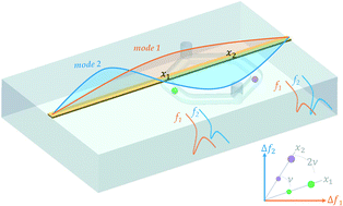
Integrated detection techniques that can characterize the morphological properties of cells are needed for the widespread use of lab-on-a-chip technology. Herein, we establish a theoretical and experimental framework to use resonant microwave sensors in their higher order modes so that the morphological properties of analytes inside a microfluidic channel can be obtained electronically. We built a phase-locked loop system that can track the first two modes of a microstrip line resonator to detect the size and location of microdroplets and cells passing through embedded microfluidic channels. The attained resolution, expressed in terms of Allan deviation at the response time, is as small as 2 × 10⁻⁸ for both modes. Additionally, simulations were performed to show that sensing with higher order modes can yield the geometrical volume, effective permittivity, two-dimensional extent, and the orientation of analytes. The framework presented here makes it possible to develop a novel type of microscope that operates at the microwave band, i.e., a radar for cells.
中文翻译:

迈向细胞的微波成像†
芯片实验室技术的广泛应用需要能够表征细胞形态特征的集成检测技术。在这里,我们建立了一个理论和实验框架,以便在其更高阶的模式下使用共振微波传感器,从而可以通过电子方式获得微流体通道内分析物的形态学特性。我们构建了一个锁相环系统,该系统可以跟踪微带线谐振器的前两种模式,以检测通过嵌入式微流体通道的微滴和细胞的大小和位置。以响应时间的艾伦偏差表示的所获得的分辨率小至2×10 -8对于两种模式。另外,进行了仿真以显示以高阶模式进行传感可以产生几何体积,有效介电常数,二维范围和分析物的方向。此处介绍的框架使开发一种在微波波段操作的新型显微镜成为可能,即用于细胞的雷达。
更新日期:2017-12-06
中文翻译:
迈向细胞的微波成像†
芯片实验室技术的广泛应用需要能够表征细胞形态特征的集成检测技术。在这里,我们建立了一个理论和实验框架,以便在其更高阶的模式下使用共振微波传感器,从而可以通过电子方式获得微流体通道内分析物的形态学特性。我们构建了一个锁相环系统,该系统可以跟踪微带线谐振器的前两种模式,以检测通过嵌入式微流体通道的微滴和细胞的大小和位置。以响应时间的艾伦偏差表示的所获得的分辨率小至2×10 -8对于两种模式。另外,进行了仿真以显示以高阶模式进行传感可以产生几何体积,有效介电常数,二维范围和分析物的方向。此处介绍的框架使开发一种在微波波段操作的新型显微镜成为可能,即用于细胞的雷达。


 京公网安备 11010802027423号