当前位置:
X-MOL 学术
›
ACS Biomater. Sci. Eng.
›
论文详情
Our official English website, www.x-mol.net, welcomes your
feedback! (Note: you will need to create a separate account there.)
Specific Nanoporous Geometries on Anodized Alumina Surfaces Influence Astrocyte Adhesion and Glial Fibrillary Acidic Protein Immunoreactivity Levels
ACS Biomaterials Science & Engineering ( IF 5.4 ) Pub Date : 2017-12-06 00:00:00 , DOI: 10.1021/acsbiomaterials.7b00760 D. Ganguly 1, 2 , C. D. L. Johnson 2, 3 , M. K. Gottipati 2, 3, 4 , D. Rende 5 , D.-A. Borca-Tasciuc 1, 6 , R. J. Gilbert 2, 3, 6
ACS Biomaterials Science & Engineering ( IF 5.4 ) Pub Date : 2017-12-06 00:00:00 , DOI: 10.1021/acsbiomaterials.7b00760 D. Ganguly 1, 2 , C. D. L. Johnson 2, 3 , M. K. Gottipati 2, 3, 4 , D. Rende 5 , D.-A. Borca-Tasciuc 1, 6 , R. J. Gilbert 2, 3, 6
Affiliation
|
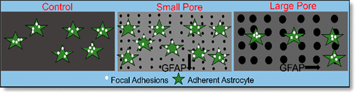
Electrodes implanted in the brain or spinal cord trigger the activation of resident astrocytes. In their reactive state, astrocytes surrounding the electrode form a glial scar, compromising the ability of the electrode to interface with the surrounding neural tissue. One approach to reduce the inhibiting scar tissue is to incorporate nanoarchitecture on the surface of the implanted materials to modify the astrocytic response. The incorporated nanoarchitecture changes both the surface characteristics and the material properties of the implant interface. We investigated the response of rat cortical astrocytes to nanoporous anodic aluminum oxide (AAO) surfaces. Astrocytes were seeded onto nonporous aluminum control surfaces and AAO surfaces with average nanopore diameters of 20 and 90 nm. The surfaces were characterized by assessing their nanomorphology, hydrophobicity, surface chemistry, mechanical properties, and surface roughness. For cell response characterization, calcein-based viability and adhesion studies were performed. Plasmid-assisted vinculin live cell imaging was done to characterize focal adhesion number and distribution. Immunocytochemistry was used to assess glial fibrillary acidic protein (GFAP) expression. We found that astrocyte adhesion was significantly higher on small pore surfaces compared to large pore surfaces. Astrocytes produced more focal adhesions (FA) and distributed these FA peripherally when cultured on small pore samples compared to the other groups. Astrocyte GFAP expression was lower when astrocytes were cultured on surfaces with small nanopores compared to the control and large pore surfaces. These results indicate that unique surface nanoporosities influence astrocyte adhesion and subsequent cellular response. The reduction in GFAP immunoreactivity exhibited by the smaller pore surfaces can improve the long-term performance of the implanted neurodevices, thus making them credible candidates as a coating material for neural implants.
中文翻译:

阳极氧化铝表面上的特定纳米孔几何形状影响星形胶质细胞粘附和胶质原纤维酸性蛋白免疫反应性水平。
植入大脑或脊髓的电极触发了星形胶质细胞的活化。处于电极反应状态的星形胶质细胞形成神经胶质瘢痕,损害了电极与周围神经组织的接触能力。减少抑制性瘢痕组织的一种方法是在植入材料的表面掺入纳米结构,以改变星形细胞的反应。并入的纳米结构改变了植入物界面的表面特性和材料特性。我们调查了大鼠皮质星形胶质细胞对纳米多孔阳极氧化铝(AAO)表面的反应。将星形胶质细胞接种到平均纳米孔直径为20和90 nm的无孔铝质对照表面和AAO表面上。通过评估其纳米形态,疏水性,表面化学性质,机械性能和表面粗糙度来表征表面。为了表征细胞反应,进行了基于钙黄绿素的生存力和粘附性研究。进行了质粒辅助的纽蛋白活细胞成像,以表征粘着斑数目和分布。免疫细胞化学用于评估神经胶质纤维酸性蛋白(GFAP)的表达。我们发现,与大孔表面相比,在小孔表面上星形胶质细胞粘附明显更高。与其他组相比,在小孔样品上培养时,星形胶质细胞产生更多的粘着斑(FA),并且将这些FA分布在周围。当在具有小纳米孔的表面上培养星形胶质细胞时,与对照和大孔表面相比,星形胶质细胞GFAP表达较低。这些结果表明独特的表面纳米孔影响星形胶质细胞粘附和随后的细胞反应。较小的孔表面表现出的GFAP免疫反应性降低可改善植入的神经装置的长期性能,从而使其成为神经植入物涂层材料的可靠候选者。
更新日期:2017-12-06
中文翻译:
阳极氧化铝表面上的特定纳米孔几何形状影响星形胶质细胞粘附和胶质原纤维酸性蛋白免疫反应性水平。
植入大脑或脊髓的电极触发了星形胶质细胞的活化。处于电极反应状态的星形胶质细胞形成神经胶质瘢痕,损害了电极与周围神经组织的接触能力。减少抑制性瘢痕组织的一种方法是在植入材料的表面掺入纳米结构,以改变星形细胞的反应。并入的纳米结构改变了植入物界面的表面特性和材料特性。我们调查了大鼠皮质星形胶质细胞对纳米多孔阳极氧化铝(AAO)表面的反应。将星形胶质细胞接种到平均纳米孔直径为20和90 nm的无孔铝质对照表面和AAO表面上。通过评估其纳米形态,疏水性,表面化学性质,机械性能和表面粗糙度来表征表面。为了表征细胞反应,进行了基于钙黄绿素的生存力和粘附性研究。进行了质粒辅助的纽蛋白活细胞成像,以表征粘着斑数目和分布。免疫细胞化学用于评估神经胶质纤维酸性蛋白(GFAP)的表达。我们发现,与大孔表面相比,在小孔表面上星形胶质细胞粘附明显更高。与其他组相比,在小孔样品上培养时,星形胶质细胞产生更多的粘着斑(FA),并且将这些FA分布在周围。当在具有小纳米孔的表面上培养星形胶质细胞时,与对照和大孔表面相比,星形胶质细胞GFAP表达较低。这些结果表明独特的表面纳米孔影响星形胶质细胞粘附和随后的细胞反应。较小的孔表面表现出的GFAP免疫反应性降低可改善植入的神经装置的长期性能,从而使其成为神经植入物涂层材料的可靠候选者。


 京公网安备 11010802027423号