Our official English website, www.x-mol.net, welcomes your
feedback! (Note: you will need to create a separate account there.)
Uracil removal-inhibited ligase reaction in combination with catalytic hairpin assembly for the sensitive and specific detection of uracil-DNA glycosylase activity
Analyst ( IF 3.6 ) Pub Date : 2017-11-06 00:00:00 , DOI: 10.1039/c7an01666b Xiaowen Xu 1, 2, 3, 4, 5 , Lei Wang 3, 5, 6, 7 , Yushu Wu 1, 2, 3, 4, 5 , Wei Jiang 1, 2, 3, 4, 5
Analyst ( IF 3.6 ) Pub Date : 2017-11-06 00:00:00 , DOI: 10.1039/c7an01666b Xiaowen Xu 1, 2, 3, 4, 5 , Lei Wang 3, 5, 6, 7 , Yushu Wu 1, 2, 3, 4, 5 , Wei Jiang 1, 2, 3, 4, 5
Affiliation

|
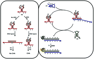
Sensitive and specific detection of uracil-DNA glycosylase (UDG) activity is crucial in biomedical study and disease diagnosis. Here, we developed a uracil removal-inhibited ligase reaction in combination with catalytic hairpin assembly (CHA) for the sensitive and specific detection of UDG activity. A hairpin probe is specially designed, which contains two uracil bases in the loop and is extended with toehold and branch-migration domains at the ends of the stem. Two short oligonucleotides are separately hybridized to one-half of the loop of the hairpin probe to form a DNA complex with a nick. Under the action of UDG, two uracil bases in the hairpin-loop are removed to generate apurinic/apyrimidinic (AP) sites. The AP sites locating at the 3′-side of the nick inhibit the ligase reaction, leaving the toehold and branch-migration domains at the ends of the hairpin probe still adjacent. The adjacent toehold and branch-migration domains initiate CHA, producing numerous G-quadruplex (G4) structures, which interact with N-methyl-mesoporphyrin IX (NMM) to generate an enhanced fluorescence signal. The excessive probes would be masked by the ligase reaction that closes the nick and forms a long DNA strand fully complementary to the hairpin domain. The probes then get opened and the toehold/branch-migration domains are not associated, prohibiting the CHA reaction and minimizing false-positive interferences. The detection limit is as low as 0.00028 U mL−1, and UDG can be well distinguished from other DNA glycosylases. Furthermore, this method is successfully applied for detecting UDG activity from HeLa cell lysates. Additionally, the inhibition of UDG activity is analyzed, which shows inhibitor dose-dependent activity suppression. This strategy will provide a promising tool for assaying UDG activity in biomedical study and disease diagnosis.
中文翻译:

尿嘧啶去除抑制连接酶反应与催化发夹装配相结合,可灵敏地检测尿嘧啶-DNA糖基化酶活性
灵敏和特异性检测尿嘧啶DNA糖基化酶(UDG)活性在生物医学研究和疾病诊断中至关重要。在这里,我们开发了尿嘧啶去除抑制的连接酶反应与催化发夹大会(CHA)结合起来,用于UDG活性的灵敏和特异检测。发夹探针经过特殊设计,在环中包含两个尿嘧啶碱基,并在茎的末端延伸了脚趾和分支迁移域。将两个短的寡核苷酸分别与发夹探针的一半环杂交,形成带有缺口的DNA复合物。在UDG的作用下,将发夹环中的两个尿嘧啶碱基去除,以生成嘌呤/两性嘧啶(AP)位点。位于缺口3'侧的AP位点会抑制连接酶反应,使发夹探针末端的脚趾和分支迁移域仍然相邻。相邻的脚趾和分支迁移域启动CHA,产生大量的G-四链体(G4)结构,这些结构与N-甲基间卟啉IX(NMM)产生增强的荧光信号。过量的探针将被封闭缺口的连接酶反应掩盖,并形成与发夹结构域完全互补的长DNA链。然后打开探针,并且脚趾/分支迁移域不关联,从而禁止CHA反应并最大程度地减少假阳性干扰。检测限低至0.00028 U mL -1,UDG可以与其他DNA糖基化酶很好地区分开。此外,该方法已成功应用于从HeLa细胞裂解物中检测UDG活性。另外,分析了对UDG活性的抑制,其显示了抑制剂剂量依赖性活性的抑制。该策略将为在生物医学研究和疾病诊断中检测UDG活性提供有希望的工具。
更新日期:2017-11-24
中文翻译:

尿嘧啶去除抑制连接酶反应与催化发夹装配相结合,可灵敏地检测尿嘧啶-DNA糖基化酶活性
灵敏和特异性检测尿嘧啶DNA糖基化酶(UDG)活性在生物医学研究和疾病诊断中至关重要。在这里,我们开发了尿嘧啶去除抑制的连接酶反应与催化发夹大会(CHA)结合起来,用于UDG活性的灵敏和特异检测。发夹探针经过特殊设计,在环中包含两个尿嘧啶碱基,并在茎的末端延伸了脚趾和分支迁移域。将两个短的寡核苷酸分别与发夹探针的一半环杂交,形成带有缺口的DNA复合物。在UDG的作用下,将发夹环中的两个尿嘧啶碱基去除,以生成嘌呤/两性嘧啶(AP)位点。位于缺口3'侧的AP位点会抑制连接酶反应,使发夹探针末端的脚趾和分支迁移域仍然相邻。相邻的脚趾和分支迁移域启动CHA,产生大量的G-四链体(G4)结构,这些结构与N-甲基间卟啉IX(NMM)产生增强的荧光信号。过量的探针将被封闭缺口的连接酶反应掩盖,并形成与发夹结构域完全互补的长DNA链。然后打开探针,并且脚趾/分支迁移域不关联,从而禁止CHA反应并最大程度地减少假阳性干扰。检测限低至0.00028 U mL -1,UDG可以与其他DNA糖基化酶很好地区分开。此外,该方法已成功应用于从HeLa细胞裂解物中检测UDG活性。另外,分析了对UDG活性的抑制,其显示了抑制剂剂量依赖性活性的抑制。该策略将为在生物医学研究和疾病诊断中检测UDG活性提供有希望的工具。


 京公网安备 11010802027423号