Our official English website, www.x-mol.net, welcomes your
feedback! (Note: you will need to create a separate account there.)
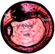
Non-staining visualization of embryogenesis and energy metabolism in medaka fish eggs using near-infrared spectroscopy and imaging
Analyst ( IF 3.6 ) Pub Date : 2017-11-23 00:00:00 , DOI: 10.1039/c7an01575e Paralee Puangchit 1, 2, 3, 4 , Mika Ishigaki 1, 2, 3, 4 , Yui Yasui 1, 2, 3, 4 , Misato Kajita 1, 2, 3, 4 , Pitiporn Ritthiruangdej 5, 6, 7, 8, 9 , Yukihiro Ozaki 1, 2, 3, 4
Analyst ( IF 3.6 ) Pub Date : 2017-11-23 00:00:00 , DOI: 10.1039/c7an01575e Paralee Puangchit 1, 2, 3, 4 , Mika Ishigaki 1, 2, 3, 4 , Yui Yasui 1, 2, 3, 4 , Misato Kajita 1, 2, 3, 4 , Pitiporn Ritthiruangdej 5, 6, 7, 8, 9 , Yukihiro Ozaki 1, 2, 3, 4
Affiliation
|
The energy metabolism and embryogenesis of fertilized Japanese medaka eggs were investigated in vivo at the molecular level using near-infrared (NIR) spectroscopy and imaging. Changes in chemical components, such as proteins and lipids, in yolk sphere and embryonic body were studied over the course of embryonic development. Metabolic changes that represent variations in the concentrations and molecular compositions of proteins and lipids in the yolk part, particularly on the 1st day after fertilization and the day just before hatching, were successfully identified in the 4900–4000 cm−1 wavenumber region. The yolk components were shown to have specific functions at the very early and final stages of the embryonic development. Proteins with α-helix- or β-sheet-rich structures clearly showed the different variation patterns within the developing egg. Furthermore, the distribution of lipids could be selectively visualized using data from the higher wavenumber region. Detailed embryonic structures were clearly depicted in the NIR images using the data from the 6400–5500 cm−1 region in which the embryo parts had some characteristic peaks due to unsaturated fatty acids. It was made clear that yolk and embryo parts had different components especially lipid components. The present study provides new insights into material variations in the fertilized egg during its growth. NIR imaging proved to be valuable in investigating the embryogenesis in vivo at the molecular level in terms of changes in biomolecular concentrations and compositions, metabolic differentiation, and detailed information about embryonic structures without the need for staining.
中文翻译:

使用近红外光谱和成像技术对红aka鱼卵的胚胎发生和能量代谢进行不染色的可视化
能量代谢和受精日本青鳉卵胚胎发生进行了研究在体内在使用近红外(NIR)光谱和成像用分子水平。在胚胎发育过程中,研究了蛋黄球和胚胎体内化学成分(例如蛋白质和脂质)的变化。表示在蛋黄部分的蛋白质和脂质的浓度的变化和分子的组合物的代谢变化,特别是在1日受精后一天和刚孵化,在4900-4000厘米成功鉴定的前一天-1波数区域。卵黄成分在胚胎发育的早期和最后阶段具有特定的功能。具有富含α-螺旋或β-折叠结构的蛋白质清楚地显示出正在发育的卵内的不同变异模式。此外,可以使用较高波数区域的数据选择性地显示脂质的分布。使用6400–5500 cm -1的数据在NIR图像中清楚地描绘了详细的胚胎结构。由于不饱和脂肪酸,其中胚胎部分具有一些特征峰的区域。已经清楚的是,蛋黄和胚胎部分具有不同的组分,特别是脂质组分。本研究为受精卵在生长过程中的物质变化提供了新的见解。在生物分子浓度和组成的变化,代谢分化以及无需染色的胚胎结构的详细信息方面,NIR成像在分子水平上研究体内胚胎发生方面被证明是有价值的。
更新日期:2017-11-23
中文翻译:
使用近红外光谱和成像技术对红aka鱼卵的胚胎发生和能量代谢进行不染色的可视化
能量代谢和受精日本青鳉卵胚胎发生进行了研究在体内在使用近红外(NIR)光谱和成像用分子水平。在胚胎发育过程中,研究了蛋黄球和胚胎体内化学成分(例如蛋白质和脂质)的变化。表示在蛋黄部分的蛋白质和脂质的浓度的变化和分子的组合物的代谢变化,特别是在1日受精后一天和刚孵化,在4900-4000厘米成功鉴定的前一天-1波数区域。卵黄成分在胚胎发育的早期和最后阶段具有特定的功能。具有富含α-螺旋或β-折叠结构的蛋白质清楚地显示出正在发育的卵内的不同变异模式。此外,可以使用较高波数区域的数据选择性地显示脂质的分布。使用6400–5500 cm -1的数据在NIR图像中清楚地描绘了详细的胚胎结构。由于不饱和脂肪酸,其中胚胎部分具有一些特征峰的区域。已经清楚的是,蛋黄和胚胎部分具有不同的组分,特别是脂质组分。本研究为受精卵在生长过程中的物质变化提供了新的见解。在生物分子浓度和组成的变化,代谢分化以及无需染色的胚胎结构的详细信息方面,NIR成像在分子水平上研究体内胚胎发生方面被证明是有价值的。


 京公网安备 11010802027423号