Our official English website, www.x-mol.net, welcomes your
feedback! (Note: you will need to create a separate account there.)
Bioconjugated fluorescent organic nanoparticles targeting EGFR-overexpressing cancer cells
Nanoscale ( IF 5.8 ) Pub Date : 2017-10-26 00:00:00 , DOI: 10.1039/c7nr06533g Adrien Faucon 1, 2, 3, 4 , Houda Benhelli-Mokrani 2, 3, 4, 5 , Fabrice Fleury 2, 3, 4, 5 , Stéphanie Dutertre 4, 6, 7, 8, 9 , Marc Tramier 4, 6, 7, 8, 9 , Joanna Boucard 1, 2, 3, 4 , Lénaïc Lartigue 1, 2, 3, 4 , Steven Nedellec 4, 10, 11 , Philippe Hulin 4, 10, 11 , Eléna Ishow 1, 2, 3, 4
Nanoscale ( IF 5.8 ) Pub Date : 2017-10-26 00:00:00 , DOI: 10.1039/c7nr06533g Adrien Faucon 1, 2, 3, 4 , Houda Benhelli-Mokrani 2, 3, 4, 5 , Fabrice Fleury 2, 3, 4, 5 , Stéphanie Dutertre 4, 6, 7, 8, 9 , Marc Tramier 4, 6, 7, 8, 9 , Joanna Boucard 1, 2, 3, 4 , Lénaïc Lartigue 1, 2, 3, 4 , Steven Nedellec 4, 10, 11 , Philippe Hulin 4, 10, 11 , Eléna Ishow 1, 2, 3, 4
Affiliation

|
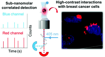
The field of optical bioimaging has considerably flourished with the advent of sophisticated microscopy techniques and ultra-bright fluorescent tools. Fluorescent organic nanoparticles (FONs) have thus recently appeared as very attractive labels for their high payload, absence of cytotoxicity and eventual biodegradation. Nevertheless, their bioconjugation to target specific receptors with high imaging contrast is scarcely performed. Moreover, assessing the reality of bioconjugation represents high challenges given the sub-nanomolar concentrations resulting from the commonly adopted nanoprecipitation fabrication process. Here, we describe how the combination of a magnetic shell allows us to easily generate red-emitting FONs conjugated with the epidermal growth factor ligand (EGF), a small protein promoting cancer cell proliferation by activating the EGF receptor (EGFR) pathway. Dual color fluorescence correlation spectroscopy combined with immunofluorescence is originally harnessed in its time trace mode to unambiguously demonstrate covalent attachment between the FON and EGF at sub-nanomolar concentrations. Strong asymmetric clustering of EGF-conjugated FONs is observed at the membrane of MDA-MB-468 human breast cancer cells overexpressing EGF receptors using super-resolution fluorescence microscopy. Such high recruitment of EGF-conjugated FONs is attributed to their EGF multivalency (4.7 EGF per FON) which enables efficient EGFR activation and subsequent phosphorylation. The large hydrodynamic diameter (DH ∼ 301 nm) of EGF-conjugated FONs prevents immediate engulfment of the sequestered receptors, which provides very bright and localized spots in less than 30 minutes. The reported bioconjugated nanoassemblies could thus serve as ultra-bright probes of breast cancer cells with EGFR-overexpression that is often associated with poor prognosis.
中文翻译:

生物共轭的荧光有机纳米粒子靶向过表达EGFR的癌细胞
随着复杂的显微镜技术和超高亮度荧光工具的出现,光学生物成像领域蓬勃发展。因此,荧光有机纳米颗粒(FON)最近因其高有效载荷,无细胞毒性和最终的生物降解而成为非常有吸引力的标记。然而,几乎没有执行它们对具有高成像对比度的靶标特异性受体的生物缀合。此外,考虑到由于通常采用的纳米沉淀制备工艺而产生的亚纳摩尔浓度,评估生物共轭的现实提出了很高的挑战。在这里,我们描述了磁壳的组合如何使我们能够轻松生成与表皮生长因子配体(EGF)共轭的发红光的FON,通过激活EGF受体(EGFR)途径促进癌细胞增殖的小蛋白。最初将双色荧光相关光谱技术与免疫荧光技术结合使用,以其时间跟踪模式明确显示了亚纳摩尔浓度时FON和EGF之间的共价结合。使用超分辨率荧光显微镜在过量表达EGF受体的MDA-MB-468人乳腺癌细胞膜上观察到EGF共轭FON的强不对称簇。如此高的EGF共轭FON募集量归因于其EGF多价(每个FON 4.7 EGF),可实现有效的EGFR激活和随后的磷酸化。大水力直径(最初将双色荧光相关光谱技术与免疫荧光技术结合使用,以其时间跟踪模式明确显示了亚纳摩尔浓度时FON和EGF之间的共价结合。使用超分辨率荧光显微镜在过量表达EGF受体的MDA-MB-468人乳腺癌细胞膜上观察到EGF共轭FON的强不对称簇。如此高的EGF共轭FON募集量归因于其EGF多价(每个FON 4.7 EGF),可实现有效的EGFR激活和随后的磷酸化。大水力直径(最初将双色荧光相关光谱技术与免疫荧光技术结合使用,以其时间跟踪模式明确显示了亚纳摩尔浓度时FON和EGF之间的共价结合。使用超分辨率荧光显微镜在过量表达EGF受体的MDA-MB-468人乳腺癌细胞膜上观察到EGF共轭FON的强烈不对称簇。如此高的EGF共轭FON募集量归因于其EGF多价(每个FON 4.7 EGF),可实现有效的EGFR激活和随后的磷酸化。大水力直径(使用超分辨率荧光显微镜在过量表达EGF受体的MDA-MB-468人乳腺癌细胞膜上观察到EGF共轭FON的强不对称簇。如此高的EGF共轭FON募集量归因于其EGF多价(每个FON 4.7 EGF),可实现有效的EGFR激活和随后的磷酸化。大水力直径(使用超分辨率荧光显微镜在过量表达EGF受体的MDA-MB-468人乳腺癌细胞膜上观察到EGF共轭FON的强不对称簇。如此高的EGF共轭FON募集量归因于其EGF多价(每个FON 4.7 EGF),可实现有效的EGFR激活和随后的磷酸化。大水力直径(d ħ的〜301 nm)的EGF-缀合FONS防止隔离受体的即时吞没,它提供了在小于30分钟非常明亮和局部位置。因此,已报道的生物共轭纳米组装体可作为乳腺癌细胞超亮探针,其EGFR过表达通常与不良预后有关。
更新日期:2017-11-23
中文翻译:

生物共轭的荧光有机纳米粒子靶向过表达EGFR的癌细胞
随着复杂的显微镜技术和超高亮度荧光工具的出现,光学生物成像领域蓬勃发展。因此,荧光有机纳米颗粒(FON)最近因其高有效载荷,无细胞毒性和最终的生物降解而成为非常有吸引力的标记。然而,几乎没有执行它们对具有高成像对比度的靶标特异性受体的生物缀合。此外,考虑到由于通常采用的纳米沉淀制备工艺而产生的亚纳摩尔浓度,评估生物共轭的现实提出了很高的挑战。在这里,我们描述了磁壳的组合如何使我们能够轻松生成与表皮生长因子配体(EGF)共轭的发红光的FON,通过激活EGF受体(EGFR)途径促进癌细胞增殖的小蛋白。最初将双色荧光相关光谱技术与免疫荧光技术结合使用,以其时间跟踪模式明确显示了亚纳摩尔浓度时FON和EGF之间的共价结合。使用超分辨率荧光显微镜在过量表达EGF受体的MDA-MB-468人乳腺癌细胞膜上观察到EGF共轭FON的强不对称簇。如此高的EGF共轭FON募集量归因于其EGF多价(每个FON 4.7 EGF),可实现有效的EGFR激活和随后的磷酸化。大水力直径(最初将双色荧光相关光谱技术与免疫荧光技术结合使用,以其时间跟踪模式明确显示了亚纳摩尔浓度时FON和EGF之间的共价结合。使用超分辨率荧光显微镜在过量表达EGF受体的MDA-MB-468人乳腺癌细胞膜上观察到EGF共轭FON的强不对称簇。如此高的EGF共轭FON募集量归因于其EGF多价(每个FON 4.7 EGF),可实现有效的EGFR激活和随后的磷酸化。大水力直径(最初将双色荧光相关光谱技术与免疫荧光技术结合使用,以其时间跟踪模式明确显示了亚纳摩尔浓度时FON和EGF之间的共价结合。使用超分辨率荧光显微镜在过量表达EGF受体的MDA-MB-468人乳腺癌细胞膜上观察到EGF共轭FON的强烈不对称簇。如此高的EGF共轭FON募集量归因于其EGF多价(每个FON 4.7 EGF),可实现有效的EGFR激活和随后的磷酸化。大水力直径(使用超分辨率荧光显微镜在过量表达EGF受体的MDA-MB-468人乳腺癌细胞膜上观察到EGF共轭FON的强不对称簇。如此高的EGF共轭FON募集量归因于其EGF多价(每个FON 4.7 EGF),可实现有效的EGFR激活和随后的磷酸化。大水力直径(使用超分辨率荧光显微镜在过量表达EGF受体的MDA-MB-468人乳腺癌细胞膜上观察到EGF共轭FON的强不对称簇。如此高的EGF共轭FON募集量归因于其EGF多价(每个FON 4.7 EGF),可实现有效的EGFR激活和随后的磷酸化。大水力直径(d ħ的〜301 nm)的EGF-缀合FONS防止隔离受体的即时吞没,它提供了在小于30分钟非常明亮和局部位置。因此,已报道的生物共轭纳米组装体可作为乳腺癌细胞超亮探针,其EGFR过表达通常与不良预后有关。


 京公网安备 11010802027423号