当前位置:
X-MOL 学术
›
Adv. Funct. Mater.
›
论文详情
Our official English website, www.x-mol.net, welcomes your
feedback! (Note: you will need to create a separate account there.)
Colorectal Cancer Diagnosis Using Enzyme‐Sensitive Ratiometric Fluorescence Dye and Antibody–Quantum Dot Conjugates for Multiplexed Detection
Advanced Functional Materials ( IF 18.5 ) Pub Date : 2017-11-22 , DOI: 10.1002/adfm.201703450 Youngrong Park 1 , Yeon-Mi Ryu 2 , Taejun Wang 3 , Yebin Jung 1 , Sohee Kim 1 , Sekyu Hwang 1 , Joonhyuck Park 1 , Dong-Jun Bae 2 , Jaeil Kim 4 , Heejo Moon 1 , Hyun-Suk Lim 1 , Sang-Yeob Kim 2, 5 , Euiheon Chung 6 , Ki Hean Kim 3, 7 , Sungjee Kim 1, 8 , Seung-Jae Myung 2, 9
Advanced Functional Materials ( IF 18.5 ) Pub Date : 2017-11-22 , DOI: 10.1002/adfm.201703450 Youngrong Park 1 , Yeon-Mi Ryu 2 , Taejun Wang 3 , Yebin Jung 1 , Sohee Kim 1 , Sekyu Hwang 1 , Joonhyuck Park 1 , Dong-Jun Bae 2 , Jaeil Kim 4 , Heejo Moon 1 , Hyun-Suk Lim 1 , Sang-Yeob Kim 2, 5 , Euiheon Chung 6 , Ki Hean Kim 3, 7 , Sungjee Kim 1, 8 , Seung-Jae Myung 2, 9
Affiliation
|
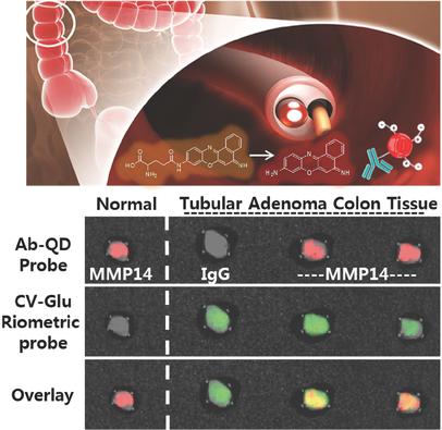
A rapid and accurate molecular fluorescence imaging technique will greatly reduce cancer mortality by overcoming the detection limit of the naked eye in colonoscopy. Two imaging probes are reported that can be co‐used for colonoscopic diagnosis: a fluorescent molecular probe, cresyl violet–glutamic acid derivative, that ratiometrically switches between two fluorescent colors in response to the enzyme activity of λ‐glutamyltranspeptidase and an antibody quantum dot probe that is a conjugate of biocompatible AgInS2 quantum dot with matrix metalloproteinase 14 antibodies. Validity of the probes is confirmed using human colon cancer cell lines, ex vivo mouse model tissues, and patient tumor colon tissues in which the tumor lesions are well‐visualized in less than five minutes. Co‐application of the two probes onto fresh colon tissues affords accurate visualization of carcinomas and also hyperplasia and adenoma regions. Fresh human colon adenoma tissues are also valuated, where the two probes show complementary diagnoses of cancer. Two‐photon microscopy shows the time‐dependent depth profiles of the two probes. Both rapidly permeate and populate most at 10–20 µm from the surface. Extensive toxicity studies are performed for the two probes at cellular level and also at the organ level using a small animal model.
中文翻译:

酶敏比例荧光染料和抗体-量子点结合物对大肠癌的多重检测
快速准确的分子荧光成像技术将克服结肠镜检查中肉眼的检测极限,从而大大降低癌症死亡率。据报道,有两种成像探针可以共同用于结肠镜诊断:一种荧光分子探针,甲酚紫-谷氨酸衍生物,响应于λ-谷氨酰转肽酶的酶活性和一种抗体量子点探针,可按比例在两种荧光色之间切换这是生物相容性AgInS 2的结合物带有基质金属蛋白酶14抗体的量子点。使用人类结肠癌细胞系,离体小鼠模型组织和患者肿瘤结肠组织(在不到五分钟的时间内即可清晰看到肿瘤病变)证实了探针的有效性。将两种探针共同应用到新鲜的结肠组织上,可以准确显示癌变以及增生和腺瘤区域。还评估了新鲜的人类结肠腺瘤组织,其中两种探针显示出对癌症的补充诊断。双光子显微镜显示了两个探针随时间变化的深度曲线。两者都迅速渗透并在距离表面10–20 µm处居多。使用小型动物模型对两种探针在细胞水平和器官水平进行了广泛的毒性研究。
更新日期:2017-11-22
中文翻译:
酶敏比例荧光染料和抗体-量子点结合物对大肠癌的多重检测
快速准确的分子荧光成像技术将克服结肠镜检查中肉眼的检测极限,从而大大降低癌症死亡率。据报道,有两种成像探针可以共同用于结肠镜诊断:一种荧光分子探针,甲酚紫-谷氨酸衍生物,响应于λ-谷氨酰转肽酶的酶活性和一种抗体量子点探针,可按比例在两种荧光色之间切换这是生物相容性AgInS 2的结合物带有基质金属蛋白酶14抗体的量子点。使用人类结肠癌细胞系,离体小鼠模型组织和患者肿瘤结肠组织(在不到五分钟的时间内即可清晰看到肿瘤病变)证实了探针的有效性。将两种探针共同应用到新鲜的结肠组织上,可以准确显示癌变以及增生和腺瘤区域。还评估了新鲜的人类结肠腺瘤组织,其中两种探针显示出对癌症的补充诊断。双光子显微镜显示了两个探针随时间变化的深度曲线。两者都迅速渗透并在距离表面10–20 µm处居多。使用小型动物模型对两种探针在细胞水平和器官水平进行了广泛的毒性研究。


 京公网安备 11010802027423号