Our official English website, www.x-mol.net, welcomes your
feedback! (Note: you will need to create a separate account there.)
OCT imaging detection of brain blood vessels in mouse, based on semiconducting polymer nanoparticles
Analyst ( IF 3.6 ) Pub Date : 2017-10-17 00:00:00 , DOI: 10.1039/c7an01245d Shaozhuang Yang 1, 2, 3, 4 , Haobin Chen 4, 5, 6, 7 , Liwei Liu 1, 2, 3, 4 , Bingling Chen 1, 2, 3, 4 , Zhigang Yang 1, 2, 3, 4 , Changfeng Wu 4, 5, 6, 7 , Siyi Hu 4, 8, 9, 10 , Huiyun Lin 4, 11, 12, 13, 14 , Buhong Li 4, 11, 12, 13, 14 , Junle Qu 1, 2, 3, 4
Analyst ( IF 3.6 ) Pub Date : 2017-10-17 00:00:00 , DOI: 10.1039/c7an01245d Shaozhuang Yang 1, 2, 3, 4 , Haobin Chen 4, 5, 6, 7 , Liwei Liu 1, 2, 3, 4 , Bingling Chen 1, 2, 3, 4 , Zhigang Yang 1, 2, 3, 4 , Changfeng Wu 4, 5, 6, 7 , Siyi Hu 4, 8, 9, 10 , Huiyun Lin 4, 11, 12, 13, 14 , Buhong Li 4, 11, 12, 13, 14 , Junle Qu 1, 2, 3, 4
Affiliation

|
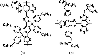
Optical Coherence Tomography (OCT) is a valuable technology that has been used to obtain microstructure images of tissue, and has several advantages, though its applications are limited in high-scattering tissues. Therefore, semiconducting polymer nanoparticles (SPNs) that possess strong absorption characteristics are applied to decrease light scattering in tissues and used as exogenous contrast agents for enhancing the contrast of OCT imaging detection. In this paper, we prepared two kinds of SPNs, termed PIDT-TBZ SPNs and PBDT-TBZ SPNs, as the contrast agents for OCT detection to enhance the signal. Firstly, we proved that they were good contrast agents for OCT imaging in agar–TiO2. After that, the contrast effects of these two SPNs were quantitatively analyzed, and then cerebral blood vessels were monitored by a home-made SD-OCT system. Finally, we created OCT images in vitro and in vivo with these two probes and performed quantitative analysis using the images. The results indicated that these SPNs created a clear contrast enhancement of small vessels in the OCT imaging process, which provides a basis for the application of SPNs as contrast agents for bioimaging studies.
中文翻译:

基于半导体高分子纳米粒子的OCT成像检测小鼠脑血管
光学相干断层扫描(OCT)是一种有价值的技术,已用于获取组织的微结构图像,尽管它在高散射组织中的应用受到限制,但它具有许多优势。因此,具有强吸收特性的半导体聚合物纳米颗粒(SPN)被用于减少组织中的光散射,并用作外源性造影剂以增强OCT成像检测的对比度。在本文中,我们准备了两种SPN,称为PIDT-TBZ SPN和PBDT-TBZ SPN,作为用于OCT检测以增强信号的造影剂。首先,我们证明了它们是琼脂– TiO 2中OCT成像的良好对比剂。。之后,定量分析这两个SPN的对比效果,然后通过自制的SD-OCT系统监测脑血管。最后,我们使用这两种探针在体外和体内创建了OCT图像,并使用这些图像进行了定量分析。结果表明,这些SPN在OCT成像过程中明显增强了小血管的对比度,这为SPN用作生物成像研究的造影剂提供了基础。
更新日期:2017-11-20
中文翻译:

基于半导体高分子纳米粒子的OCT成像检测小鼠脑血管
光学相干断层扫描(OCT)是一种有价值的技术,已用于获取组织的微结构图像,尽管它在高散射组织中的应用受到限制,但它具有许多优势。因此,具有强吸收特性的半导体聚合物纳米颗粒(SPN)被用于减少组织中的光散射,并用作外源性造影剂以增强OCT成像检测的对比度。在本文中,我们准备了两种SPN,称为PIDT-TBZ SPN和PBDT-TBZ SPN,作为用于OCT检测以增强信号的造影剂。首先,我们证明了它们是琼脂– TiO 2中OCT成像的良好对比剂。。之后,定量分析这两个SPN的对比效果,然后通过自制的SD-OCT系统监测脑血管。最后,我们使用这两种探针在体外和体内创建了OCT图像,并使用这些图像进行了定量分析。结果表明,这些SPN在OCT成像过程中明显增强了小血管的对比度,这为SPN用作生物成像研究的造影剂提供了基础。


 京公网安备 11010802027423号