Our official English website, www.x-mol.net, welcomes your
feedback! (Note: you will need to create a separate account there.)
Development of a label-free Raman imaging technique for differentiation of malaria parasite infected from non-infected tissue†
Analyst ( IF 3.6 ) Pub Date : 2017-11-16 00:00:00 , DOI: 10.1039/c7an01760j Laura Frame 1, 2, 3, 4, 5 , James Brewer 5, 6, 7, 8 , Rebecca Lee 5, 6, 7, 8 , Karen Faulds 1, 2, 3, 4, 5 , Duncan Graham 1, 2, 3, 4, 5
Analyst ( IF 3.6 ) Pub Date : 2017-11-16 00:00:00 , DOI: 10.1039/c7an01760j Laura Frame 1, 2, 3, 4, 5 , James Brewer 5, 6, 7, 8 , Rebecca Lee 5, 6, 7, 8 , Karen Faulds 1, 2, 3, 4, 5 , Duncan Graham 1, 2, 3, 4, 5
Affiliation
|
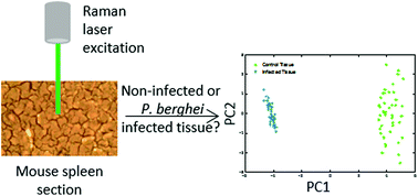
During malarial infection, the host uses the spleen to clear the malaria parasites, however, the parasites have evolved the ability to bind to endothelial receptors in blood vessels of tissues to avoid removal, known as sequestration, and this is largely responsible for the symptoms and severity of infection. So a technique which could non-invasively diagnose tissue burden could be utilised as an aid for localised malaria diagnosis within tissue. Raman spectroscopy is a label-free imaging technique and can provide unique and chemically specific Raman ‘fingerprint’ spectrum of biological samples such as tissue. Within this study, Raman imaging was used to observe the changes to the molecular composition of mice spleen tissue under malarial infection, compared with non-infected samples. From analysis of the Raman imaging data, both tissue types showed very similar spectral profiles, which highlighted that their biochemical compositions were closely linked. Principal component analysis showed very clear separation of the two sample groups, with an associated increase in concentration of heme-based Raman vibrations within the infected dataset. This was indicative of the presence of hemozoin, the malaria pigment, being detected within the infected spleen. Separation also showed that as the hemozoin content within the tissue increased, there was a corresponding change to hemoglobin and some lipid/nucleic acid vibrations. These results demonstrate that Raman spectroscopy can be used to easily discriminate the subtle changes in tissue burden upon malarial infection.
中文翻译:

开发无标记拉曼成像技术,以区分未感染组织感染的疟原虫†
在疟疾感染期间,宿主利用脾脏清除疟疾寄生虫,但是,这些寄生虫已进化出与组织血管中的内皮受体结合的能力,可以避免清除,称为螯合,这是造成症状和疾病的主要原因。感染的严重程度。因此可以利用非侵入性诊断组织负担的技术作为组织内局部疟疾诊断的辅助手段。拉曼光谱法是一种无标记的成像技术,可以提供生物样品(例如组织)的独特且化学特有的拉曼“指纹”光谱。在这项研究中,与未感染的样本相比,使用拉曼成像观察了在疟疾感染下小鼠脾脏组织的分子组成的变化。通过对拉曼成像数据的分析,两种组织类型均显示出非常相似的光谱图,这突出表明它们的生化组成密切相关。主成分分析表明,两个样本组的分离非常清晰,并且感染数据集中基于血红素的拉曼振动的浓度相应增加。这表明在感染的脾脏中检测到了疟疾色素hemozoin的存在。分离还显示,随着组织中血红蛋白含量的增加,血红蛋白发生相应变化,并且脂质/核酸发生一些振动。这些结果表明,拉曼光谱法可用于容易地区分疟疾感染后组织负担的细微变化。主成分分析表明,两个样本组的分离非常清晰,并且感染数据集中基于血红素的拉曼振动的浓度相应增加。这表明在感染的脾脏中检测到了疟疾色素hemozoin的存在。分离还显示,随着组织中血红蛋白含量的增加,血红蛋白发生相应变化,并且出现一些脂质/核酸振动。这些结果表明,拉曼光谱法可用于容易地区分疟疾感染后组织负担的细微变化。主成分分析表明,两个样本组的分离非常清晰,并且感染数据集中基于血红素的拉曼振动的浓度相应增加。这表明在感染的脾脏中检测到了疟疾色素hemozoin的存在。分离还显示,随着组织中血红蛋白含量的增加,血红蛋白发生相应变化,并且脂质/核酸发生一些振动。这些结果表明,拉曼光谱法可用于容易地区分疟疾感染后组织负担的细微变化。被感染的脾脏中检测到的疟疾色素。分离还显示,随着组织中血红蛋白含量的增加,血红蛋白发生相应变化,并且脂质/核酸发生一些振动。这些结果表明,拉曼光谱法可用于容易地区分疟疾感染后组织负担的细微变化。被感染的脾脏中检测到的疟疾色素。分离还显示,随着组织中血红蛋白含量的增加,血红蛋白发生相应变化,并且脂质/核酸发生一些振动。这些结果表明,拉曼光谱法可用于容易地区分疟疾感染后组织负担的细微变化。
更新日期:2017-11-16
中文翻译:
开发无标记拉曼成像技术,以区分未感染组织感染的疟原虫†
在疟疾感染期间,宿主利用脾脏清除疟疾寄生虫,但是,这些寄生虫已进化出与组织血管中的内皮受体结合的能力,可以避免清除,称为螯合,这是造成症状和疾病的主要原因。感染的严重程度。因此可以利用非侵入性诊断组织负担的技术作为组织内局部疟疾诊断的辅助手段。拉曼光谱法是一种无标记的成像技术,可以提供生物样品(例如组织)的独特且化学特有的拉曼“指纹”光谱。在这项研究中,与未感染的样本相比,使用拉曼成像观察了在疟疾感染下小鼠脾脏组织的分子组成的变化。通过对拉曼成像数据的分析,两种组织类型均显示出非常相似的光谱图,这突出表明它们的生化组成密切相关。主成分分析表明,两个样本组的分离非常清晰,并且感染数据集中基于血红素的拉曼振动的浓度相应增加。这表明在感染的脾脏中检测到了疟疾色素hemozoin的存在。分离还显示,随着组织中血红蛋白含量的增加,血红蛋白发生相应变化,并且脂质/核酸发生一些振动。这些结果表明,拉曼光谱法可用于容易地区分疟疾感染后组织负担的细微变化。主成分分析表明,两个样本组的分离非常清晰,并且感染数据集中基于血红素的拉曼振动的浓度相应增加。这表明在感染的脾脏中检测到了疟疾色素hemozoin的存在。分离还显示,随着组织中血红蛋白含量的增加,血红蛋白发生相应变化,并且出现一些脂质/核酸振动。这些结果表明,拉曼光谱法可用于容易地区分疟疾感染后组织负担的细微变化。主成分分析表明,两个样本组的分离非常清晰,并且感染数据集中基于血红素的拉曼振动的浓度相应增加。这表明在感染的脾脏中检测到了疟疾色素hemozoin的存在。分离还显示,随着组织中血红蛋白含量的增加,血红蛋白发生相应变化,并且脂质/核酸发生一些振动。这些结果表明,拉曼光谱法可用于容易地区分疟疾感染后组织负担的细微变化。被感染的脾脏中检测到的疟疾色素。分离还显示,随着组织中血红蛋白含量的增加,血红蛋白发生相应变化,并且脂质/核酸发生一些振动。这些结果表明,拉曼光谱法可用于容易地区分疟疾感染后组织负担的细微变化。被感染的脾脏中检测到的疟疾色素。分离还显示,随着组织中血红蛋白含量的增加,血红蛋白发生相应变化,并且脂质/核酸发生一些振动。这些结果表明,拉曼光谱法可用于容易地区分疟疾感染后组织负担的细微变化。


 京公网安备 11010802027423号