当前位置:
X-MOL 学术
›
Dalton Trans.
›
论文详情
Our official English website, www.x-mol.net, welcomes your
feedback! (Note: you will need to create a separate account there.)
Exploring the effect of substituent in the hydrazone ligand of a family of μ-oxidodivanadium(V) hydrazone complexes on structure, DNA binding and anticancer activity
Dalton Transactions ( IF 3.5 ) Pub Date : 2017-11-01 00:00:00 , DOI: 10.1039/c7dt03585c Debashis Patra 1, 2, 3, 4, 5 , Subhabrata Paul 5, 6, 7, 8 , Indira Majumder 5, 6, 7, 8 , Nayim Sepay 5, 9, 10, 11 , Sachinath Bera 5, 9, 12, 13 , Rita Kundu 5, 6, 7, 8 , Michael G. B. Drew 9, 14, 15, 16 , Tapas Ghosh 1, 2, 3, 4, 5
Dalton Transactions ( IF 3.5 ) Pub Date : 2017-11-01 00:00:00 , DOI: 10.1039/c7dt03585c Debashis Patra 1, 2, 3, 4, 5 , Subhabrata Paul 5, 6, 7, 8 , Indira Majumder 5, 6, 7, 8 , Nayim Sepay 5, 9, 10, 11 , Sachinath Bera 5, 9, 12, 13 , Rita Kundu 5, 6, 7, 8 , Michael G. B. Drew 9, 14, 15, 16 , Tapas Ghosh 1, 2, 3, 4, 5
Affiliation
|
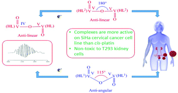
The reaction of 2-hydroxybenzoylhydrazine (H2bh) separately with equimolar amounts of [VIVO(aa)2] and [VIVO(ba)2] in CHCl3 afforded the complexes [VV2O3(HL1)2] (1) and [VV2O3(HL2)2] (2) respectively in good to excellent yield ((HL1)2− and (HL2)2− represent respectively the dianionic form of 2-hydroxybenzoylhydrazones of acetylacetone (H3L1) and benzoylacetone (H3L2) (general abbreviation H3L)). From X-ray structure analysis, the VV–O–VV angle was found to be ∼115° and 180° in 1 and 2 respectively. Upon one-electron reduction selectively at one V centre at an appropriate potential, each of 1 and 2 generated mixed-valence [(HL)VVO-(μ-O)-OVIV(HL)]− species 1A and 2A respectively, which showed valence delocalization at room temperature and localization at 77 K, and the VIV–O–VV bond angles were calculated to be 177.5° and 180° respectively. The intercalative mode of binding of the two complexes 1 and 2 with CT DNA has been suggested by UV-visible spectroscopy (Kb = 7.31 × 10⁵ M−1 and 8.71 × 10⁵ M−1 respectively for 1 and 2), fluorescence spectroscopy (Ksv = 6.85 × 10⁵ M−1 and 8.53 × 10⁵ M−1 respectively for 1 and 2) and circular dichroism spectroscopy. Such intercalative mode of binding of these two complexes with CT DNA and HPV DNA has also been confirmed by molecular docking study. Both complexes 1 and 2 exhibited promising anti-cancer activity against SiHa cervical cancer cells with IC50 values of 28 ± 0.5 μM and 25 ± 0.5 μM respectively for 24 h which is significantly better than that of widely used cisplatin (with IC50 value of 63.5 μM). Nuclear staining experiments reveal that these complexes kill the SiHa cells through apoptotic mode. It is interesting to note that these two complexes are non-toxic to normal T293 cell line. Complex 2 showed higher DNA binding ability with CT DNA and HPV DNA as well as better anti-cancer properties towards SiHa cervical cancer cells in comparison to complex 1, a fact which can be explained by considering the lower energy of LUMO (which favours electron transition from DNA to the metal complex) and also the higher surface area of complex 2 in comparison to complex 1 due to the presence of one extra electron-withdrawing phenyl group in the former.
中文翻译:

探索μ-氧化二钒(V)配合物家族的ligand配体中的取代基对结构,DNA结合和抗癌活性的影响
2-羟基苯甲酰肼(H 2 bh)分别与等摩尔量的[V IV O(aa)2 ]和[V IV O(ba)2 ]在CHCl 3中反应,得到络合物[V V 2 O 3(HL 1)2 ](1)和[V V 2 O 3(HL 2)2 ](2)分别具有良好至极好的收率((HL 1)2−和(HL 2)2−分别表示的乙酰丙酮的2- hydroxybenzoylhydrazones(H双阴离子形式3大号1)和苯甲酰丙酮(H 3大号2)(一般缩写ħ 3 L))。通过X射线结构分析,发现V V –O – V V角在1和2中分别为〜115°和180° 。在适当的电势下,在一个V中心有选择地还原一个电子时,1和2各自生成混合价[(HL)V V O-(μ-O)-OV IV(HL)] -种类1A和2A分别显示了在室温和在77 K处的化合价离域化,并且V IV –O–V V键角分别计算为177.5°和180°。的两种复合物结合的插层模式1和2与CT DNA已经建议通过UV-可见光谱(ķ b = 7.31×10 5中号-1和8.71×10 5中号-1分别为1和2),荧光光谱学(K sv = 6.85×10 5 M -1和8.53×10 5 M-1分别为1和2)和圆二色光谱。分子对接研究也证实了这两种复合物与CT DNA和HPV DNA的这种结合方式。配合物1和2均显示出对SiHa宫颈癌细胞的有希望的抗癌活性,其24小时的IC 50值分别为28±0.5μM和25±0.5μM,这显着优于广泛使用的顺铂(IC 50值为50)。 63.5μM)。核染色实验表明,这些复合物通过凋亡模式杀死SiHa细胞。有趣的是,这两种复合物对正常的T293细胞系无毒。复杂2与复合物1相比,显示出与CT DNA和HPV DNA更高的DNA结合能力以及对SiHa宫颈癌细胞的更好的抗癌特性,这一事实可以通过考虑较低的LUMO能量来解释(这有利于DNA的电子跃迁)由于络合物1中存在一个额外的吸电子苯基,因此与络合物1相比,络合物2的表面积更大。
更新日期:2017-11-15
中文翻译:
探索μ-氧化二钒(V)配合物家族的ligand配体中的取代基对结构,DNA结合和抗癌活性的影响
2-羟基苯甲酰肼(H 2 bh)分别与等摩尔量的[V IV O(aa)2 ]和[V IV O(ba)2 ]在CHCl 3中反应,得到络合物[V V 2 O 3(HL 1)2 ](1)和[V V 2 O 3(HL 2)2 ](2)分别具有良好至极好的收率((HL 1)2−和(HL 2)2−分别表示的乙酰丙酮的2- hydroxybenzoylhydrazones(H双阴离子形式3大号1)和苯甲酰丙酮(H 3大号2)(一般缩写ħ 3 L))。通过X射线结构分析,发现V V –O – V V角在1和2中分别为〜115°和180° 。在适当的电势下,在一个V中心有选择地还原一个电子时,1和2各自生成混合价[(HL)V V O-(μ-O)-OV IV(HL)] -种类1A和2A分别显示了在室温和在77 K处的化合价离域化,并且V IV –O–V V键角分别计算为177.5°和180°。的两种复合物结合的插层模式1和2与CT DNA已经建议通过UV-可见光谱(ķ b = 7.31×10 5中号-1和8.71×10 5中号-1分别为1和2),荧光光谱学(K sv = 6.85×10 5 M -1和8.53×10 5 M-1分别为1和2)和圆二色光谱。分子对接研究也证实了这两种复合物与CT DNA和HPV DNA的这种结合方式。配合物1和2均显示出对SiHa宫颈癌细胞的有希望的抗癌活性,其24小时的IC 50值分别为28±0.5μM和25±0.5μM,这显着优于广泛使用的顺铂(IC 50值为50)。 63.5μM)。核染色实验表明,这些复合物通过凋亡模式杀死SiHa细胞。有趣的是,这两种复合物对正常的T293细胞系无毒。复杂2与复合物1相比,显示出与CT DNA和HPV DNA更高的DNA结合能力以及对SiHa宫颈癌细胞的更好的抗癌特性,这一事实可以通过考虑较低的LUMO能量来解释(这有利于DNA的电子跃迁)由于络合物1中存在一个额外的吸电子苯基,因此与络合物1相比,络合物2的表面积更大。


 京公网安备 11010802027423号