Our official English website, www.x-mol.net, welcomes your
feedback! (Note: you will need to create a separate account there.)
Complementary Imaging of Silver Nanoparticle Interactions with Green Algae: Dark-Field Microscopy, Electron Microscopy, and Nanoscale Secondary Ion Mass Spectrometry
ACS Nano ( IF 15.8 ) Pub Date : 2017-11-13 00:00:00 , DOI: 10.1021/acsnano.7b04556 Ryo Sekine 1, 2 , Katie L. Moore 3, 4 , Marianne Matzke 2 , Pascal Vallotton 5, 6 , Haibo Jiang 3, 7 , Gareth M. Hughes 3 , Jason K. Kirby 7 , Erica Donner 1 , Chris R. M. Grovenor 3 , Claus Svendsen 2 , Enzo Lombi 1
ACS Nano ( IF 15.8 ) Pub Date : 2017-11-13 00:00:00 , DOI: 10.1021/acsnano.7b04556 Ryo Sekine 1, 2 , Katie L. Moore 3, 4 , Marianne Matzke 2 , Pascal Vallotton 5, 6 , Haibo Jiang 3, 7 , Gareth M. Hughes 3 , Jason K. Kirby 7 , Erica Donner 1 , Chris R. M. Grovenor 3 , Claus Svendsen 2 , Enzo Lombi 1
Affiliation

|
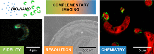
Increasing consumer use of engineered nanomaterials has led to significantly increased efforts to understand their potential impact on the environment and living organisms. Currently, no individual technique can provide all the necessary information such as their size, distribution, and chemistry in complex biological systems. Consequently, there is a need to develop complementary instrumental imaging approaches that provide enhanced understanding of these “bio-nano” interactions to overcome the limitations of individual techniques. Here we used a multimodal imaging approach incorporating dark-field light microscopy, high-resolution electron microscopy, and nanoscale secondary ion mass spectrometry (NanoSIMS). The aim was to gain insight into the bio-nano interactions of surface-functionalized silver nanoparticles (Ag-NPs) with the green algae Raphidocelis subcapitata, by combining the fidelity, spatial resolution, and elemental identification offered by the three techniques, respectively. Each technique revealed that Ag-NPs interact with the green algae with a dependence on the size (10 nm vs 60 nm) and surface functionality (tannic acid vs branched polyethylenimine, bPEI) of the NPs. Dark-field light microscopy revealed the presence of strong light scatterers on the algal cell surface, and SEM imaging confirmed their nanoparticulate nature and localization at nanoscale resolution. NanoSIMS imaging confirmed their chemical identity as Ag, with the majority of signal concentrated at the cell surface. Furthermore, SEM and NanoSIMS provided evidence of 10 nm bPEI Ag-NP internalization at higher concentrations (40 μg/L), correlating with the highest toxicity observed from these NPs. This multimodal approach thus demonstrated an effective approach to complement dose–response studies in nano-(eco)-toxicological investigations.
中文翻译:

与纳米藻相互作用的银纳米粒子相互作用的互补成像:暗场显微镜,电子显微镜和纳米级二次离子质谱。
消费者对工程纳米材料的越来越多的使用,导致人们越来越多地努力了解其对环境和生物的潜在影响。当前,在复杂的生物系统中,没有任何一种单独的技术可以提供所有必要的信息,例如它们的大小,分布和化学。因此,需要开发互补的仪器成像方法,以增强对这些“生物-纳米”相互作用的理解,以克服单个技术的局限性。在这里,我们使用了包含暗场光学显微镜,高分辨率电子显微镜和纳米级二次离子质谱(NanoSIMS)的多峰成像方法。目的是深入了解表面功能化的银纳米颗粒(Ag-NPs)与绿藻的生物-纳米相互作用Raphidocelis subcapitata,分别结合了三种技术提供的保真度,空间分辨率和元素识别。每种技术都表明,Ag-NPs与绿藻相互作用,这取决于大小(10 nm对60 nm)和表面功能(单宁酸对10 nm)。NP的支链聚乙烯亚胺(bPEI)。暗场光学显微镜显示藻类细胞表面存在强光散射体,并且SEM成像证实了它们的纳米颗粒性质和纳米级分辨率下的定位。NanoSIMS成像证实了它们与Ag的化学同一性,大部分信号都集中在细胞表面。此外,SEM和NanoSIMS提供了在较高浓度(40μg/ L)下10 nm bPEI Ag-NP内在化的证据,这与从这些NP观察到的最高毒性相关。因此,这种多峰方法证明了一种有效的方法,可以补充纳米(生态)毒理学研究中的剂量反应研究。
更新日期:2017-11-14
中文翻译:

与纳米藻相互作用的银纳米粒子相互作用的互补成像:暗场显微镜,电子显微镜和纳米级二次离子质谱。
消费者对工程纳米材料的越来越多的使用,导致人们越来越多地努力了解其对环境和生物的潜在影响。当前,在复杂的生物系统中,没有任何一种单独的技术可以提供所有必要的信息,例如它们的大小,分布和化学。因此,需要开发互补的仪器成像方法,以增强对这些“生物-纳米”相互作用的理解,以克服单个技术的局限性。在这里,我们使用了包含暗场光学显微镜,高分辨率电子显微镜和纳米级二次离子质谱(NanoSIMS)的多峰成像方法。目的是深入了解表面功能化的银纳米颗粒(Ag-NPs)与绿藻的生物-纳米相互作用Raphidocelis subcapitata,分别结合了三种技术提供的保真度,空间分辨率和元素识别。每种技术都表明,Ag-NPs与绿藻相互作用,这取决于大小(10 nm对60 nm)和表面功能(单宁酸对10 nm)。NP的支链聚乙烯亚胺(bPEI)。暗场光学显微镜显示藻类细胞表面存在强光散射体,并且SEM成像证实了它们的纳米颗粒性质和纳米级分辨率下的定位。NanoSIMS成像证实了它们与Ag的化学同一性,大部分信号都集中在细胞表面。此外,SEM和NanoSIMS提供了在较高浓度(40μg/ L)下10 nm bPEI Ag-NP内在化的证据,这与从这些NP观察到的最高毒性相关。因此,这种多峰方法证明了一种有效的方法,可以补充纳米(生态)毒理学研究中的剂量反应研究。


 京公网安备 11010802027423号