JACC: Cardiovascular Imaging ( IF 12.8 ) Pub Date : 2017-11-01 , DOI: 10.1016/j.jcmg.2016.10.007 Calvin W L Chin 1 , Russell J Everett 2 , Jacek Kwiecinski 3 , Alex T Vesey 2 , Emily Yeung 2 , Gavin Esson 2 , William Jenkins 2 , Maria Koo 2 , Saeed Mirsadraee 2 , Audrey C White 2 , Alan G Japp 2 , Sanjay K Prasad 4 , Scott Semple 5 , David E Newby 2 , Marc R Dweck 2

|
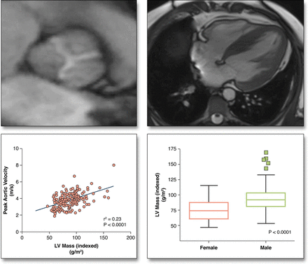
Objectives Cardiac magnetic resonance (CMR) was used to investigate the extracellular compartment and myocardial fibrosis in patients with aortic stenosis, as well as their association with other measures of left ventricular decompensation and mortality.
Background Progressive myocardial fibrosis drives the transition from hypertrophy to heart failure in aortic stenosis. Diffuse fibrosis is associated with extracellular volume expansion that is detectable by T1 mapping, whereas late gadolinium enhancement (LGE) detects replacement fibrosis.
Methods In a prospective observational cohort study, 203 subjects (166 with aortic stenosis [69 years; 69% male]; 37 healthy volunteers [68 years; 65% male]) underwent comprehensive phenotypic characterization with clinical imaging and biomarker evaluation. On CMR, we quantified the total extracellular volume of the myocardium indexed to body surface area (iECV). The iECV upper limit of normal from the control group (22.5 ml/m2) was used to define extracellular compartment expansion. Areas of replacement mid-wall LGE were also identified. All-cause mortality was determined during 2.9 ± 0.8 years of follow up.
Results iECV demonstrated a good correlation with diffuse histological fibrosis on myocardial biopsies (r = 0.87; p < 0.001; n = 11) and was increased in patients with aortic stenosis (23.6 ± 7.2 ml/m2 vs. 16.1 ± 3.2 ml/m2 in control subjects; p < 0.001). iECV was used together with LGE to categorize patients with normal myocardium (iECV <22.5 ml/m2; 51% of patients), extracellular expansion (iECV ≥22.5 ml/m2; 22%), and replacement fibrosis (presence of mid-wall LGE, 27%). There was evidence of increasing hypertrophy, myocardial injury, diastolic dysfunction, and longitudinal systolic dysfunction consistent with progressive left ventricular decompensation (all p < 0.05) across these groups. Moreover, this categorization was of prognostic value with stepwise increases in unadjusted all-cause mortality (8 deaths/1,000 patient-years vs. 36 deaths/1,000 patient-years vs. 71 deaths/1,000 patient-years, respectively; p = 0.009).
Conclusions CMR detects ventricular decompensation in aortic stenosis through the identification of myocardial extracellular expansion and replacement fibrosis. This holds major promise in tracking myocardial health in valve disease and for optimizing the timing of valve replacement. (The Role of Myocardial Fibrosis in Patients With Aortic Stenosis; NCT01755936)
中文翻译:

主动脉瓣狭窄的心肌纤维化和心脏失代偿
目的心脏磁共振 (CMR) 用于研究主动脉瓣狭窄患者的细胞外室和心肌纤维化,以及它们与左心室失代偿和死亡率其他指标的关系。
背景进行性心肌纤维化驱动主动脉瓣从肥大向心力衰竭的转变。弥漫性纤维化与 T1 标测可检测到的细胞外体积扩张有关,而晚期钆增强 (LGE) 可检测到替代纤维化。
方法在一项前瞻性观察性队列研究中,203 名受试者(166 名主动脉瓣狭窄 [69 岁;69% 男性];37 名健康志愿者 [68 岁;65% 男性])通过临床影像学和生物标志物评估进行了全面的表型表征。在 CMR 上,我们量化了与体表面积 (iECV) 相关的心肌细胞外总体积。对照组的 iECV 正常上限 (22.5 ml/m 2 ) 用于定义细胞外隔室扩张。还确定了更换中墙 LGE 的区域。在 2.9 ± 0.8 年的随访期间确定全因死亡率。
结果iECV 在心肌活检中显示出与弥漫性组织学纤维化的良好相关性(r = 0.87;p < 0.001;n = 11)并且在主动脉瓣狭窄患者中增加(23.6 ± 7.2 ml/m 2 vs. 16.1 ± 3.2 ml/m 2对照组;p < 0.001)。iECV 与 LGE 一起用于对心肌正常(iECV <22.5 ml/m 2;51% 的患者)、细胞外扩张(iECV ≥22.5 ml/m 2; 22%)和替代性纤维化(存在中壁 LGE,27%)。有证据表明,这些组的肥大、心肌损伤、舒张功能障碍和纵向收缩功能障碍与进行性左心室失代偿相一致(所有 p < 0.05)。此外,该分类具有预后价值,未调整的全因死亡率逐步增加(分别为 8 例死亡/1,000 患者年对 36 例死亡/1,000 患者年对 71 例死亡/1,000 患者年;p = 0.009) .
结论CMR通过识别心肌细胞外扩张和替代纤维化来检测主动脉瓣狭窄的心室失代偿。这在跟踪瓣膜疾病中的心肌健康和优化瓣膜置换的时机方面具有重要意义。(心肌纤维化在主动脉瓣狭窄患者中的作用;NCT01755936)


 京公网安备 11010802027423号