当前位置:
X-MOL 学术
›
Toxicol. Res.
›
论文详情
Our official English website, www.x-mol.net, welcomes your
feedback! (Note: you will need to create a separate account there.)
Involvement of histone hypoacetylation in INH-induced rat liver injury
Toxicology Research ( IF 2.2 ) Pub Date : 2017-10-04 00:00:00 , DOI: 10.1039/c7tx00166e Ling-yan Zhu 1, 2, 3, 4, 5 , Qi Ren 1, 2, 3, 4, 5 , Yu-hong Li 1, 2, 3, 4, 5 , Yi-yang Zhang 1, 2, 3, 4, 5 , Jin-feng Li 1, 2, 3, 4, 5 , Ying-shu Li 1, 2, 3, 4, 5 , Zhe Shi 1, 2, 3, 4, 5 , Fu-min Feng 1, 2, 3, 4, 5
Toxicology Research ( IF 2.2 ) Pub Date : 2017-10-04 00:00:00 , DOI: 10.1039/c7tx00166e Ling-yan Zhu 1, 2, 3, 4, 5 , Qi Ren 1, 2, 3, 4, 5 , Yu-hong Li 1, 2, 3, 4, 5 , Yi-yang Zhang 1, 2, 3, 4, 5 , Jin-feng Li 1, 2, 3, 4, 5 , Ying-shu Li 1, 2, 3, 4, 5 , Zhe Shi 1, 2, 3, 4, 5 , Fu-min Feng 1, 2, 3, 4, 5
Affiliation

|
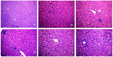
This study explores the mechanism of histone acetylation under the effect of oxidative stress in rat liver injury induced by isoniazid (INH). Fifty-six adult SD rats were selected and divided randomly into INH groups (48) and control (8). Rats in INH groups were intragastrically injected with 55 mg kg−1 day−1 for 3, 7, 10, 14, 21, and 28 days, and control rats were given an equal volume of distilled water. Pathological changes in liver tissues were observed by HE staining. Western blot analysis was conducted to measure the expression levels of H3k14ac and H4k8ac. The activities of HAT, HDAC and IL-1β, and TNF-α were detected by ELISA in liver tissues. Real-time RT-PCR analysis was performed to determine the protein expression levels of HAT, HDAC, and IL-1β and the mRNA expression of TNF-α. The levels of superoxide dismutase (SOD) and malondialdehyde (MDA) were assayed by biochemical methods in liver tissues. At different time points, the SOD activity decreased, whereas the MDA content significantly increased after 14 days (FSOD = 11.15, FMDA = 7.42, P < 0.01). During this period, the expression of histone acetylated H3K14 and H4K8 acetylation decreased compared with the control group (FH3K14 = 4.18, FH4K8 = 3.87, P < 0.05); by contrast, HDAC1 and HDAC2 showed a high expression level compared with those in the control group (FHDAC1 = 29.13, FHDAC2 = 58.34, P < 0.01). Moreover, the expression of CBP/P300 was lower than that in the control group (FCBP/P300 = 12.18, P = 0.001), and the protein contents of IL-1β and TNF-α in rat liver tissues were up-regulated (FIL-1β = 44.88, FTNF-α = 41.56, P < 0.01). These results suggest that histone acetylation is involved in INH-induced rat liver injury. Furthermore, the hypoacetylation of histones H3K14 and H4K8 is negatively correlated with oxidative stress-mediated rat liver injury.
中文翻译:

组蛋白低乙酰化参与INH诱导的大鼠肝损伤
本研究探讨了在异烟肼(INH)诱导的大鼠肝损伤中氧化应激作用下组蛋白乙酰化的机制。选择五十六只成年SD大鼠,并随机分为INH组(48只)和对照组(8只)。INH组的大鼠胃内注射55 mg kg -1天-1持续3、7、10、14、21和28天,并给对照组大鼠等量的蒸馏水。通过HE染色观察肝组织的病理变化。进行蛋白质印迹分析以测量H3k14ac和H4k8ac的表达水平。ELISA法检测肝组织中HAT,HDAC,IL-1β和TNF-α的活性。进行实时RT-PCR分析以确定HAT,HDAC和IL-1β的蛋白表达水平以及TNF-α的mRNA表达。用生化方法测定肝组织中的超氧化物歧化酶(SOD)和丙二醛(MDA)的水平。14天后,SOD活性在不同时间点下降,而MDA含量显着增加(F SOD = 11.15,F MDA = 7.42,P <0.01)。在此期间,组蛋白乙酰化的H3K14和H4K8乙酰化的表达与对照组相比有所降低(F H3K14 = 4.18,F H4K8 = 3.87,P <0.05)。相比之下,与对照组相比,HDAC1和HDAC2的表达水平较高(F HDAC1 = 29.13,F HDAC2 = 58.34,P <0.01)。此外,CBP / P300的表达低于对照组(F CBP / P300 = 12.18,P = 0.001),并且大鼠肝组织中IL-1β和TNF-α的蛋白质含量上调(˚F IL-1β= 44.88,˚F TNF-α = 41.56,P <0.01)。这些结果表明,组蛋白乙酰化与INH诱导的大鼠肝损伤有关。此外,组蛋白H3K14和H4K8的过乙酰化与氧化应激介导的大鼠肝损伤呈负相关。
更新日期:2017-11-02
中文翻译:

组蛋白低乙酰化参与INH诱导的大鼠肝损伤
本研究探讨了在异烟肼(INH)诱导的大鼠肝损伤中氧化应激作用下组蛋白乙酰化的机制。选择五十六只成年SD大鼠,并随机分为INH组(48只)和对照组(8只)。INH组的大鼠胃内注射55 mg kg -1天-1持续3、7、10、14、21和28天,并给对照组大鼠等量的蒸馏水。通过HE染色观察肝组织的病理变化。进行蛋白质印迹分析以测量H3k14ac和H4k8ac的表达水平。ELISA法检测肝组织中HAT,HDAC,IL-1β和TNF-α的活性。进行实时RT-PCR分析以确定HAT,HDAC和IL-1β的蛋白表达水平以及TNF-α的mRNA表达。用生化方法测定肝组织中的超氧化物歧化酶(SOD)和丙二醛(MDA)的水平。14天后,SOD活性在不同时间点下降,而MDA含量显着增加(F SOD = 11.15,F MDA = 7.42,P <0.01)。在此期间,组蛋白乙酰化的H3K14和H4K8乙酰化的表达与对照组相比有所降低(F H3K14 = 4.18,F H4K8 = 3.87,P <0.05)。相比之下,与对照组相比,HDAC1和HDAC2的表达水平较高(F HDAC1 = 29.13,F HDAC2 = 58.34,P <0.01)。此外,CBP / P300的表达低于对照组(F CBP / P300 = 12.18,P = 0.001),并且大鼠肝组织中IL-1β和TNF-α的蛋白质含量上调(˚F IL-1β= 44.88,˚F TNF-α = 41.56,P <0.01)。这些结果表明,组蛋白乙酰化与INH诱导的大鼠肝损伤有关。此外,组蛋白H3K14和H4K8的过乙酰化与氧化应激介导的大鼠肝损伤呈负相关。


 京公网安备 11010802027423号