JAMA Psychiatry ( IF 22.5 ) Pub Date : 2017-11-01 , DOI: 10.1001/jamapsychiatry.2017.2663 Stefan P Brugger 1, 2, 3 , Oliver D Howes 2, 3, 4
|
Importance Schizophrenia is associated with alterations in mean regional brain volumes. However, it is not known whether the clinical heterogeneity seen in the disorder is reflected at the neurobiological level, for example, in differences in the interindividual variability of these brain volumes relative to control individuals.
Objective To investigate whether patients with first-episode schizophrenia exhibit greater variability of regional brain volumes in addition to mean volume differences.
Data Sources Studies that reported regional brain volumetric measures in patients and controls by using magnetic resonance imaging in the MEDLINE, EMBASE, and PsycINFO databases from inception to October 1, 2016, were examined.
Study Selection Case-control studies that reported regional brain volumes in patients with first-episode schizophrenia and healthy controls by using magnetic resonance imaging were selected.
Data Extraction and Synthesis Means and variances (SDs) were extracted for each measure to calculate effect sizes, which were combined using multivariate meta-analysis.
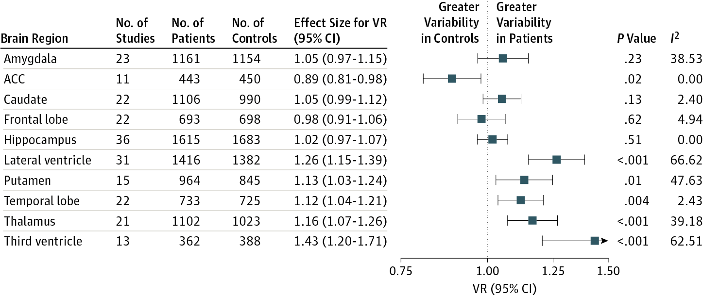
Main Outcomes and Measures Relative variability of regional brain volumetric measurements in patients compared with control groups as indexed by the variability ratio (VR) and coefficient of variation ratio (CVR). Hedges g was used to quantify mean differences.
Results A total of 108 studies that reported measurements from 3901 patients (1272 [32.6%] female) with first-episode schizophrenia and 4040 controls (1613 [39.9%] female) were included in the analyses. Variability of putamen (VR, 1.13; 95% CI, 1.03-1.24; P = .01), temporal lobe (VR, 1.12; 95% CI, 1.04-1.21; P = .004), thalamus (VR, 1.16; 95% CI, 1.07-1.26; P < .001), and third ventricle (VR, 1.43; 95% CI, 1.20-1.71; P < 1 × 10⁻⁵) volume was significantly greater in patients, whereas variability of anterior cingulate cortex volume was lower (VR, 0.89; 95% CI, 0.81-0.98; P = .02). These findings were robust to choice of outcome measure. There was no evidence of altered variability of caudate nucleus or frontal lobe volumes. Mean volumes of the lateral (g = 0.40; 95% CI, 0.29-0.51; P < .001) and third ventricles (g = 0.43; 95% CI, 0.26-0.59; P < .001) were greater, whereas mean volumes of the amygdala (g = −0.46; −0.65 to −0.26; P < .001), anterior cingulate cortex (g = −0.26; 95% CI, −0.43 to −0.10; P = .005), frontal lobe (g = −0.31; 95% CI, −0.44 to −0.19; P = .001), hippocampus (g = −0.66; 95% CI, −0.84 to −0.47; P < .001), temporal lobe (g = −0.22; 95% CI, −0.36 to −0.09; P = .001), and thalamus (g = −0.36; 95% CI, −0.57 to −0.15; P = .001) were lower in patients. There was no evidence of altered mean volume of caudate nucleus or putamen.
Conclusions and Relevance In addition to altered mean volume of many brain structures, schizophrenia is associated with significantly greater variability of temporal cortex, thalamus, putamen, and third ventricle volumes, consistent with biological heterogeneity in these regions, but lower variability of anterior cingulate cortex volume. This finding indicates greater homogeneity of anterior cingulate volume and, considered with the significantly lower mean volume of this region, suggests that this is a core region affected by the disorder.
中文翻译:
精神分裂症区域脑结构的异质性和同质性Meta分析
重要性 精神分裂症与平均区域脑容量的改变有关。然而,尚不清楚该疾病中所见的临床异质性是否反映在神经生物学水平,例如,这些脑容量相对于对照个体的个体间变异性差异。
目的 探讨首发精神分裂症患者除了平均体积差异外,是否表现出更大的区域脑容量变异性。
数据来源 研究报告了从开始到 2016 年 10 月 1 日在 MEDLINE、EMBASE 和 PsycINFO 数据库中使用磁共振成像对患者和对照组进行区域脑容量测量的研究。
研究选择 通过使用磁共振成像报告首发精神分裂症患者和健康对照的区域脑容量的病例对照研究被选中。
数据提取和综合 均值和方差 (SDs) 被提取为每个测量计算效应大小,使用多元荟萃分析结合。
主要结果和措施 与对照组相比,患者区域脑容量测量的相对变异性,以变异比 (VR) 和变异系数比 (CVR) 为指标。对冲g用于量化平均差异。
结果 共有 108 项研究报告了 3901 名首发精神分裂症患者(1272 名 [32.6%] 女性)和 4040 名对照者(1613 名 [39.9%] 女性)的测量结果,这些结果被纳入分析。壳核的变异性 (VR, 1.13; 95% CI, 1.03-1.24; P = .01)、颞叶 (VR, 1.12; 95% CI, 1.04-1.21; P = .004)、丘脑 (VR, 1.16; 95 % CI, 1.07-1.26; P < .001) 和第三脑室 (VR, 1.43; 95% CI, 1.20-1.71; P < 1 × 10 -5 ) 体积在患者中显着增加,而前扣带皮层的变异性体积较低(VR,0.89;95% CI,0.81-0.98;P = .02)。这些发现对于结果测量的选择是稳健的。没有证据表明尾状核或额叶体积的变异性发生改变。侧脑室(g = 0.40;95% CI,0.29-0.51;P < .001)和第三脑室(g = 0.43;95% CI,0.26-0.59;P < .001)的平均体积更大,而平均体积更大杏仁核 ( g = -0.46; -0.65 至 -0.26; P < .001),前扣带皮层 ( g = -0.26; 95% CI, -0.43 至 -0.10; P = .005),额叶 ( g = -0.31;95% CI,-0.44 至 -0.19;P = .001),海马(g = -0.66;95% CI,-0.84 至 -0.47;P < .001)、颞叶(g = -0.22;95% CI,-0.36 至 -0.09;P = .001)和丘脑(g = -0.36;95% CI,-0.57 至 -0.15;P = .001)在患者中较低。没有证据表明尾状核或壳核的平均体积发生改变。
结论和相关性 除了许多大脑结构的平均体积改变外,精神分裂症与颞叶皮层、丘脑、壳核和第三脑室体积的变异性显着增加相关,这与这些区域的生物异质性一致,但前扣带皮层体积的变异性较低. 这一发现表明前扣带回体积更均匀,并且考虑到该区域的平均体积显着降低,表明这是受疾病影响的核心区域。


 京公网安备 11010802027423号