当前位置:
X-MOL 学术
›
J. Hepatol.
›
论文详情
Our official English website, www.x-mol.net, welcomes your
feedback! (Note: you will need to create a separate account there.)
Evaluation of HCC Response to Locoregional Therapy: Validation of MRI-Based Response Criteria versus Explant Pathology
Journal of Hepatology ( IF 26.8 ) Pub Date : 2017-12-01 , DOI: 10.1016/j.jhep.2017.07.030 Sonja Gordic , Idoia Corcuera-Solano , Ashley Stueck , Cecilia Besa , Pamela Argiriadi , Preethi Guniganti , Michael King , Shingo Kihira , James Babb , Swan Thung , Bachir Taouli
Journal of Hepatology ( IF 26.8 ) Pub Date : 2017-12-01 , DOI: 10.1016/j.jhep.2017.07.030 Sonja Gordic , Idoia Corcuera-Solano , Ashley Stueck , Cecilia Besa , Pamela Argiriadi , Preethi Guniganti , Michael King , Shingo Kihira , James Babb , Swan Thung , Bachir Taouli
|
BACKGROUND AND AIMS
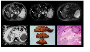
This study evaluates the performance of various magnetic resonance imaging (MRI) response criteria for the prediction of complete pathologic necrosis (CPN) of hepatocellular carcinoma (HCC) post locoregional therapy (LRT) using explant pathology as a reference. METHODS
We included 61 patients (male/female 46/15; mean age 60years) who underwent liver transplantation after LRT with transarterial chemoembolization plus radiofrequency or microwave ablation (n=56), or 90Yttrium radioembolization (n=5). MRI was performed <90days before liver transplantation. Three independent readers assessed the following criteria: RECIST, EASL, modified RECIST (mRECIST), percentage of necrosis on subtraction images, and diffusion-weighted imaging (DWI), both qualitative (signal intensity) and quantitative (apparent diffusion coefficient [ADC]). The degree of necrosis was retrospectively assessed at histopathology. Intraclass correlation coefficient (ICC) and Cohen's kappa were used to assess inter-reader agreement. Logistic regression and receiver operating characteristic analyses were used to determine imaging predictors of CPN. Pearson correlation was performed between imaging criteria and pathologic degree of tumor necrosis. RESULTS
A total of 97HCCs (mean size 2.3±1.3cm) including 28 with CPN were evaluated. There was excellent inter-reader agreement (ICC 0.77-0.86, all methods). EASL, mRECIST, percentage of necrosis and qualitative DWI were all significant (p<0.001) predictors of CPN, while RECIST and ADC were not. EASL, mRECIST and percentage of necrosis performed similarly (area under the curves [AUCs] 0.810-0.815) while the performance of qualitative DWI was lower (AUC 0.622). Image subtraction demonstrated the strongest correlation (r=0.71-0.72, p<0.0001) with pathologic degree of tumor necrosis. CONCLUSIONS
EASL/mRECIST criteria and image subtraction have excellent diagnostic performance for predicting CPN in HCC treated with LRT, with image subtraction correlating best with pathologic degree of tumor necrosis. Thus, MR image subtraction is recommended for assessing HCC response to LRT. LAY SUMMARY
The assessment of hepatocellular carcinoma (HCC) tumor necrosis after locoregional therapy is essential for additional treatment planning and estimation of outcome. In this study, we assessed the performance of various magnetic resonance imaging (MRI) response criteria (RECIST, mRECIST, EASL, percentage of necrosis on subtraction images, and diffusion-weighted imaging) for the prediction of complete pathologic necrosis of HCC post locoregional therapy on liver explant. Patients who underwent liver transplantation after locoregional therapy were included in this retrospective study. All patients underwent routine liver MRI within 90days of liver transplantation. EASL/mRECIST criteria and image subtraction had excellent diagnostic performance for predicting complete pathologic necrosis in treated HCC, with image subtraction correlating best with pathologic degree of tumor necrosis.
中文翻译:

评估 HCC 对局部治疗的反应:基于 MRI 的反应标准与外植体病理学的验证
背景和目的 本研究使用外植体病理学作为参考,评估了各种磁共振成像 (MRI) 反应标准在预测局部区域治疗 (LRT) 后肝细胞癌 (HCC) 完全病理性坏死 (CPN) 方面的性能。方法 我们纳入了 61 名患者(男性/女性 46/15;平均年龄 60 岁),他们在 LRT 后接受肝移植,经动脉化疗栓塞加射频或微波消融术(n=56),或 90 钇放射栓塞术(n=5)。MRI 在肝移植前 <90 天进行。三位独立的读者评估了以下标准:RECIST、EASL、改良 RECIST (mRECIST)、减影图像上的坏死百分比和扩散加权成像 (DWI),包括定性(信号强度)和定量(表观扩散系数 [ADC]) . 在组织病理学上回顾性评估坏死的程度。组内相关系数 (ICC) 和 Cohen's kappa 用于评估读者间的一致性。Logistic 回归和接收器操作特征分析用于确定 CPN 的成像预测因子。影像学标准与肿瘤坏死的病理程度进行Pearson相关。结果 共评估了 97 个 HCC(平均大小 2.3±1.3cm),其中 28 个患有 CPN。读者之间的一致性非常好(ICC 0.77-0.86,所有方法)。EASL、mRECIST、坏死百分比和定性 DWI 都是 CPN 的显着预测因子 (p<0.001),而 RECIST 和 ADC 则不是。EASL、mRECIST 和坏死百分比表现相似(曲线下面积 [AUC] 0.810-0.815),而定性 DWI 的表现较低(AUC 0. 622)。图像减影显示与肿瘤坏死的病理程度最强的相关性(r=0.71-0.72,p<0.0001)。结论 EASL/mRECIST 标准和图像减影对于预测 LRT 治疗的 HCC 的 CPN 具有良好的诊断性能,图像减影与肿瘤坏死的病理程度相关性最好。因此,推荐使用 MR 图像减影来评估 HCC 对 LRT 的反应。概述 局部治疗后肝细胞癌 (HCC) 肿瘤坏死的评估对于额外的治疗计划和结果评估至关重要。在这项研究中,我们评估了各种磁共振成像 (MRI) 反应标准(RECIST、mRECIST、EASL、减影图像上的坏死百分比、和扩散加权成像)用于预测肝外植体局部治疗后 HCC 的完全病理性坏死。本回顾性研究包括在局部治疗后接受肝移植的患者。所有患者在肝移植后 90 天内均接受了常规肝脏 MRI 检查。EASL/mRECIST 标准和图像减影对于预测已治疗 HCC 的完全病理性坏死具有出色的诊断性能,图像减影与肿瘤坏死的病理程度相关性最好。
更新日期:2017-12-01
中文翻译:
评估 HCC 对局部治疗的反应:基于 MRI 的反应标准与外植体病理学的验证
背景和目的 本研究使用外植体病理学作为参考,评估了各种磁共振成像 (MRI) 反应标准在预测局部区域治疗 (LRT) 后肝细胞癌 (HCC) 完全病理性坏死 (CPN) 方面的性能。方法 我们纳入了 61 名患者(男性/女性 46/15;平均年龄 60 岁),他们在 LRT 后接受肝移植,经动脉化疗栓塞加射频或微波消融术(n=56),或 90 钇放射栓塞术(n=5)。MRI 在肝移植前 <90 天进行。三位独立的读者评估了以下标准:RECIST、EASL、改良 RECIST (mRECIST)、减影图像上的坏死百分比和扩散加权成像 (DWI),包括定性(信号强度)和定量(表观扩散系数 [ADC]) . 在组织病理学上回顾性评估坏死的程度。组内相关系数 (ICC) 和 Cohen's kappa 用于评估读者间的一致性。Logistic 回归和接收器操作特征分析用于确定 CPN 的成像预测因子。影像学标准与肿瘤坏死的病理程度进行Pearson相关。结果 共评估了 97 个 HCC(平均大小 2.3±1.3cm),其中 28 个患有 CPN。读者之间的一致性非常好(ICC 0.77-0.86,所有方法)。EASL、mRECIST、坏死百分比和定性 DWI 都是 CPN 的显着预测因子 (p<0.001),而 RECIST 和 ADC 则不是。EASL、mRECIST 和坏死百分比表现相似(曲线下面积 [AUC] 0.810-0.815),而定性 DWI 的表现较低(AUC 0. 622)。图像减影显示与肿瘤坏死的病理程度最强的相关性(r=0.71-0.72,p<0.0001)。结论 EASL/mRECIST 标准和图像减影对于预测 LRT 治疗的 HCC 的 CPN 具有良好的诊断性能,图像减影与肿瘤坏死的病理程度相关性最好。因此,推荐使用 MR 图像减影来评估 HCC 对 LRT 的反应。概述 局部治疗后肝细胞癌 (HCC) 肿瘤坏死的评估对于额外的治疗计划和结果评估至关重要。在这项研究中,我们评估了各种磁共振成像 (MRI) 反应标准(RECIST、mRECIST、EASL、减影图像上的坏死百分比、和扩散加权成像)用于预测肝外植体局部治疗后 HCC 的完全病理性坏死。本回顾性研究包括在局部治疗后接受肝移植的患者。所有患者在肝移植后 90 天内均接受了常规肝脏 MRI 检查。EASL/mRECIST 标准和图像减影对于预测已治疗 HCC 的完全病理性坏死具有出色的诊断性能,图像减影与肿瘤坏死的病理程度相关性最好。


 京公网安备 11010802027423号