当前位置:
X-MOL 学术
›
J. Mater. Chem. B
›
论文详情
Our official English website, www.x-mol.net, welcomes your feedback! (Note: you will need to create a separate account there.)
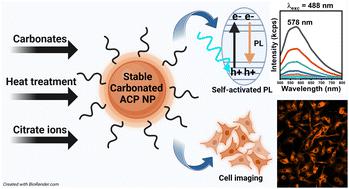
Tailoring the structure and self-activated photoluminescence of carbonated amorphous calcium phosphate nanoparticles for bioimaging applications
Journal of Materials Chemistry B ( IF 7 ) Pub Date : 2024-04-18 , DOI: 10.1039/d3tb02915h Thales R. Machado 1 , Carlos E. Zanardo 1 , Raquel R.C. Vilela 1 , Renata R. Miranda 1 , Natália S. Moreno 1 , Celisnolia M. Leite 1 , Elson Longo 2 , Valtencir Zucolotto 1
Journal of Materials Chemistry B ( IF 7 ) Pub Date : 2024-04-18 , DOI: 10.1039/d3tb02915h Thales R. Machado 1 , Carlos E. Zanardo 1 , Raquel R.C. Vilela 1 , Renata R. Miranda 1 , Natália S. Moreno 1 , Celisnolia M. Leite 1 , Elson Longo 2 , Valtencir Zucolotto 1
Affiliation
|
Self-activated luminescent calcium phosphate (CaP) nanoparticles, including hydroxyapatite (HA) and amorphous calcium phosphate (ACP), are promising for bioimaging and theragnostic applications in nanomedicine, eliminating the need for activator ions or fluorophores. In this study, we developed luminescent and stable citrate-functionalized carbonated ACP nanoparticles for bioimaging purposes. Our findings revealed that both the CO32− content and the posterior heating step at 400 °C significantly influenced the composition and the structural ordering of the chemically precipitated ACP nanoparticles, impacting the intensity, broadness, and position of the defect-related photoluminescence (PL) emission band. The heat-treated samples also exhibited excitation-dependent PL under excitation wavelengths typically used in bioimaging (λexc = 405, 488, 561, and 640 nm). Citrate functionalization improved the PL intensity of the nanoparticles by inhibiting non-radiative deactivation mechanisms in solution. Additionally, it resulted in an increased colloidal stability and reduced aggregation, high stability of the metastable amorphous phase and the PL emission for at least 96 h in water and supplemented culture medium. MTT assay of HepaRG cells, incubated for 24 and 48 h with the nanoparticles in concentrations ranging from 10 to 320 μg mL−1, evidenced their high biocompatibility. Internalization studies using the nanoparticles self-activated luminescence showed that cellular uptake of the nanoparticles is both time (4–24 h) and concentration (160–320 μg mL−1) dependent. Experiments using confocal laser scanning microscopy allowed the successful imaging of the nanoparticles inside cells via their intrinsic PL after 4 h of incubation. Our results highlight the potential use of citrate-functionalized carbonated ACP nanoparticles for use in internalization assays and bioimaging procedures.
中文翻译:

定制用于生物成像应用的碳酸化无定形磷酸钙纳米颗粒的结构和自激活光致发光
自激活发光磷酸钙 (CaP) 纳米粒子,包括羟基磷灰石 (HA) 和无定形磷酸钙 (ACP),有望用于纳米医学中的生物成像和治疗诊断应用,从而消除对激活剂离子或荧光团的需求。在这项研究中,我们开发了用于生物成像目的的发光且稳定的柠檬酸官能化碳酸 ACP 纳米颗粒。我们的研究结果表明,CO 3 2−含量和 400 °C 的后加热步骤显着影响化学沉淀 ACP 纳米粒子的组成和结构排序,影响与缺陷相关的光致发光的强度、宽度和位置( PL) 发射带。热处理的样品还在生物成像中常用的激发波长(λ exc = 405、488、561 和 640 nm)下表现出与激发相关的 PL。柠檬酸盐官能化通过抑制溶液中的非辐射失活机制来提高纳米粒子的PL强度。此外,它还导致胶体稳定性增加、聚集减少、亚稳态非晶相的高稳定性以及在水和补充培养基中至少 96 小时的 PL 发射。用浓度范围为10至320 μg mL -1的纳米颗粒孵育24和48小时的HepaRG细胞的MTT测定证明了它们的高生物相容性。使用纳米粒子自激活发光的内化研究表明,纳米粒子的细胞摄取既依赖于时间(4-24 小时)又依赖于浓度(160-320 μg mL -1)。使用共焦激光扫描显微镜进行的实验允许在孵育 4 小时后通过其内在 PL成功对细胞内的纳米颗粒进行成像。我们的结果强调了柠檬酸盐功能化的碳酸 ACP 纳米颗粒在内化测定和生物成像过程中的潜在用途。
更新日期:2024-04-18
中文翻译:
定制用于生物成像应用的碳酸化无定形磷酸钙纳米颗粒的结构和自激活光致发光
自激活发光磷酸钙 (CaP) 纳米粒子,包括羟基磷灰石 (HA) 和无定形磷酸钙 (ACP),有望用于纳米医学中的生物成像和治疗诊断应用,从而消除对激活剂离子或荧光团的需求。在这项研究中,我们开发了用于生物成像目的的发光且稳定的柠檬酸官能化碳酸 ACP 纳米颗粒。我们的研究结果表明,CO 3 2−含量和 400 °C 的后加热步骤显着影响化学沉淀 ACP 纳米粒子的组成和结构排序,影响与缺陷相关的光致发光的强度、宽度和位置( PL) 发射带。热处理的样品还在生物成像中常用的激发波长(λ exc = 405、488、561 和 640 nm)下表现出与激发相关的 PL。柠檬酸盐官能化通过抑制溶液中的非辐射失活机制来提高纳米粒子的PL强度。此外,它还导致胶体稳定性增加、聚集减少、亚稳态非晶相的高稳定性以及在水和补充培养基中至少 96 小时的 PL 发射。用浓度范围为10至320 μg mL -1的纳米颗粒孵育24和48小时的HepaRG细胞的MTT测定证明了它们的高生物相容性。使用纳米粒子自激活发光的内化研究表明,纳米粒子的细胞摄取既依赖于时间(4-24 小时)又依赖于浓度(160-320 μg mL -1)。使用共焦激光扫描显微镜进行的实验允许在孵育 4 小时后通过其内在 PL成功对细胞内的纳米颗粒进行成像。我们的结果强调了柠檬酸盐功能化的碳酸 ACP 纳米颗粒在内化测定和生物成像过程中的潜在用途。


 京公网安备 11010802027423号