当前位置:
X-MOL 学术
›
Ind. Eng. Chem. Res.
›
论文详情
Our official English website, www.x-mol.net, welcomes your feedback! (Note: you will need to create a separate account there.)
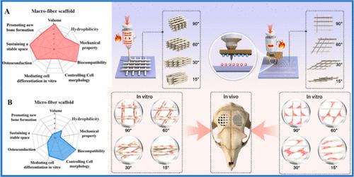
Macro- or Microfiber Scaffolds with Different Fiber Arrangements: Determining the Best Design for Osteogenesis
Industrial & Engineering Chemistry Research ( IF 4.2 ) Pub Date : 2024-04-16 , DOI: 10.1021/acs.iecr.3c03934 Xiangjun Yang 1, 2 , He Cai 1 , Ruyi Li 1 , Zihan He 1 , Yanhua Liu 1 , Jiaxin Wu 1 , Zhengyi Xu 1 , Linxin Yang 1 , Zhou Zhu 1 , Jian Wang 1 , Xibo Pei 1 , Junyu Chen 1 , Qianbing Wan 1
Industrial & Engineering Chemistry Research ( IF 4.2 ) Pub Date : 2024-04-16 , DOI: 10.1021/acs.iecr.3c03934 Xiangjun Yang 1, 2 , He Cai 1 , Ruyi Li 1 , Zihan He 1 , Yanhua Liu 1 , Jiaxin Wu 1 , Zhengyi Xu 1 , Linxin Yang 1 , Zhou Zhu 1 , Jian Wang 1 , Xibo Pei 1 , Junyu Chen 1 , Qianbing Wan 1
Affiliation
|
Bone tissue engineering scaffolds with varied 3D structures have been investigated extensively. The arrangements and diameters of scaffold fibers are usually considered during the scaffold manufacturing processes. There is still controversy over the optimal angle of arrangement and fiber diameter of scaffolds for bone tissue engineering. To highlight the problem, we herein designed macrofiber (diameter: 551.3 ± 22.11 μm) and microfiber (diameter: 10.8 ± 0.85 μm) scaffolds with fibers arranged at 90, 60, 30, and 15°. Physicochemical characterization revealed that macrofiber scaffolds had better mechanical properties and hydrophilicity than microfiber ones. Although the different fiber arrangements of the macrofiber scaffold showed no significant differences in alkaline phosphatase expression and calcium nodule deposition, the macrofiber scaffold enhanced new bone formation obviously in the skull defect model. The microfiber scaffolds with deposition angles of 90 and 60° efficiently promoted cell osteogenic differentiation by regulating the YAP pathway and enhancing the expression of Runx2 and OCN. However, microfiber scaffolds seemed to be a barrier to new bone formation when applied for bone tissue repair of skull defects. Poor mechanical properties severely limited the biological application of microfiber scaffolds. The macrostructures guaranteed sufficient mechanical strength for osteogenic space maintenance. For clinical application, macrofiber scaffolds with high mechanical strength might be a promising therapy for repairing large bone tissue defects.
中文翻译:

具有不同纤维排列的粗纤维或超细纤维支架:确定成骨的最佳设计
具有不同 3D 结构的骨组织工程支架已得到广泛研究。在支架制造过程中通常要考虑支架纤维的排列和直径。对于骨组织工程支架的最佳排列角度和纤维直径仍存在争议。为了突出这个问题,我们设计了粗纤维(直径:551.3±22.11μm)和微纤维(直径:10.8±0.85μm)支架,纤维排列为90°、60°、30°和15°。物理化学表征表明,粗纤维支架比微纤维支架具有更好的机械性能和亲水性。尽管粗纤维支架的不同纤维排列在碱性磷酸酶表达和钙结节沉积方面没有显着差异,但在颅骨缺损模型中,粗纤维支架明显增强了新骨形成。沉积角为90°和60°的微纤维支架通过调节YAP通路、增强Runx2和OCN的表达,有效促进细胞成骨分化。然而,当应用于颅骨缺损的骨组织修复时,微纤维支架似乎成为新骨形成的障碍。机械性能差严重限制了微纤维支架的生物学应用。宏观结构保证了成骨空间维持足够的机械强度。对于临床应用,具有高机械强度的粗纤维支架可能是修复大骨组织缺损的有前途的疗法。
更新日期:2024-04-17
中文翻译:
具有不同纤维排列的粗纤维或超细纤维支架:确定成骨的最佳设计
具有不同 3D 结构的骨组织工程支架已得到广泛研究。在支架制造过程中通常要考虑支架纤维的排列和直径。对于骨组织工程支架的最佳排列角度和纤维直径仍存在争议。为了突出这个问题,我们设计了粗纤维(直径:551.3±22.11μm)和微纤维(直径:10.8±0.85μm)支架,纤维排列为90°、60°、30°和15°。物理化学表征表明,粗纤维支架比微纤维支架具有更好的机械性能和亲水性。尽管粗纤维支架的不同纤维排列在碱性磷酸酶表达和钙结节沉积方面没有显着差异,但在颅骨缺损模型中,粗纤维支架明显增强了新骨形成。沉积角为90°和60°的微纤维支架通过调节YAP通路、增强Runx2和OCN的表达,有效促进细胞成骨分化。然而,当应用于颅骨缺损的骨组织修复时,微纤维支架似乎成为新骨形成的障碍。机械性能差严重限制了微纤维支架的生物学应用。宏观结构保证了成骨空间维持足够的机械强度。对于临床应用,具有高机械强度的粗纤维支架可能是修复大骨组织缺损的有前途的疗法。


 京公网安备 11010802027423号