Nature Protocols ( IF 14.8 ) Pub Date : 2024-04-11 , DOI: 10.1038/s41596-024-00983-3 Ying Chen , Yiwei Yang , Fan Zhang
|
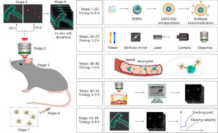
In vivo microscopy of single cells enables following pathological changes in tissues, revealing signaling networks and cell interactions critical to disease progression. However, conventional intravital microscopy at visible and near-infrared wavelengths <900 nm (NIR-I) suffers from attenuation and is typically performed following the surgical creation of an imaging window. Such surgical procedures cause the alteration of the local vasculature and induce inflammation in skin, muscle and skull, inevitably altering the microenvironment in the imaging area. Here, we detail the use of near-infrared fluorescence (NIR-II, 1,000–1,700 nm) for in vivo microscopy to circumvent attenuation in living tissues. This approach enables the noninvasive visualization of cell migration in deep tissues by labeling specific cells with NIR-II lanthanide downshifting nanoparticles exhibiting high physicochemical stability and photostability. We further developed a NIR-II fluorescence microscopy setup for in vivo imaging through the intact skull with high spatiotemporal resolution, which we use for the real-time dynamic visualization of single-neutrophil behavior in the deep brain of a mouse model of ischemic stroke. The labeled downshifting nanoparticle synthesis takes 5–6 d, the imaging system setup takes 1–2 h, the in vivo cell labeling takes 1–3 h, the in vivo NIR-II microscopic imaging takes 3–5 h and the data analysis takes 3–8 h. The procedures can be performed by users with standard laboratory training in nanomaterials research and appropriate animal handling.
中文翻译:
通过 NIR-II 荧光纳米材料对小鼠大脑中的单个中性粒细胞进行无创体内显微镜观察
单细胞的体内显微镜可以跟踪组织的病理变化,揭示对疾病进展至关重要的信号网络和细胞相互作用。然而,可见光和近红外波长<900 nm (NIR-I) 的传统活体显微镜会受到衰减的影响,并且通常在手术创建成像窗口后进行。此类外科手术会导致局部脉管系统的改变,并诱发皮肤、肌肉和颅骨炎症,不可避免地改变成像区域的微环境。在这里,我们详细介绍了近红外荧光(NIR-II,1,000–1,700 nm)在活体显微镜中的使用,以避免活组织中的衰减。该方法通过用具有高物理化学稳定性和光稳定性的 NIR-II 镧系元素降频纳米粒子标记特定细胞,实现深层组织中细胞迁移的无创可视化。我们进一步开发了一种 NIR-II 荧光显微镜装置,用于通过具有高时空分辨率的完整头骨进行体内成像,我们将其用于缺血性中风小鼠模型深部大脑中单个中性粒细胞行为的实时动态可视化。标记的降频纳米颗粒合成需要 5-6 天,成像系统设置需要 1-2 小时,体内细胞标记需要 1-3 小时,体内 NIR-II 显微成像需要 3-5 小时,数据分析需要3-8 小时。这些程序可由接受过纳米材料研究和适当动物处理方面标准实验室培训的用户执行。


 京公网安备 11010802027423号