当前位置:
X-MOL 学术
›
Nano Lett.
›
论文详情
Our official English website, www.x-mol.net, welcomes your feedback! (Note: you will need to create a separate account there.)
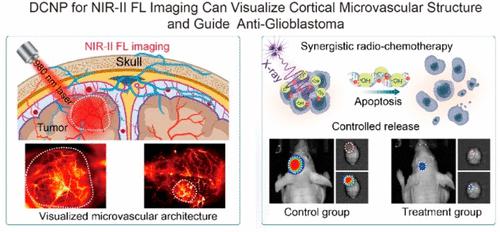
X-ray Activated Nanoprodrug for Visualization of Cortical Microvascular Alterations and NIR-II Image-Guided Chemo-Radiotherapy of Glioblastoma
Nano Letters ( IF 10.8 ) Pub Date : 2024-03-18 , DOI: 10.1021/acs.nanolett.4c00223 Lichao Su 1 , Kang Zhu 2 , Xiaoguang Ge 1 , Ying Wu 2 , Jieping Zhang 3 , Guoyu Wang 3 , Daojia Liu 3 , Ling Chen 4 , Qingqing Li 1 , Junqiang Chen 3 , Jibin Song 2
Nano Letters ( IF 10.8 ) Pub Date : 2024-03-18 , DOI: 10.1021/acs.nanolett.4c00223 Lichao Su 1 , Kang Zhu 2 , Xiaoguang Ge 1 , Ying Wu 2 , Jieping Zhang 3 , Guoyu Wang 3 , Daojia Liu 3 , Ling Chen 4 , Qingqing Li 1 , Junqiang Chen 3 , Jibin Song 2
Affiliation
|
The permeability of the highly selective blood–brain barrier (BBB) to anticancer drugs and the difficulties in defining deep tumor boundaries often reduce the effectiveness of glioma treatment. Thus, exploring the combination of multiple treatment modalities under the guidance of second-generation near-infrared (NIR-II) window fluorescence (FL) imaging is considered a strategic approach in glioma theranostics. Herein, a hybrid X-ray-activated nanoprodrug was developed to precisely visualize the structural features of glioma microvasculature and delineate the boundary of glioma for synergistic chemo-radiotherapy. The nanoprodrug comprised down-converted nanoparticle (DCNP) coated with X-ray sensitive poly(Se-Se/DOX-co-acrylic acid) and targeted Angiopep-2 peptide (DCNP@P(Se-DOX)@ANG). Because of its ultrasmall size and the presence of DOX, the nanoprodrug could easily cross BBB to precisely monitor and localize glioblastoma via intracranial NIR-II FL imaging and synergistically administer antiglioblastoma chemo-radiotherapy through specific X-ray-induced DOX release and radiosensitization. This study provides a novel and effective strategy for glioblastoma imaging and chemo-radiotherapy.
中文翻译:

X 射线激活纳米前药用于皮质微血管变化的可视化和 NIR-II 图像引导胶质母细胞瘤的化疗放疗
高度选择性血脑屏障(BBB)对抗癌药物的渗透性以及确定肿瘤深部边界的困难通常会降低神经胶质瘤治疗的有效性。因此,在第二代近红外(NIR-II)窗口荧光(FL)成像指导下探索多种治疗方式的组合被认为是胶质瘤治疗诊断的战略方法。在此,开发了一种混合 X 射线激活纳米前药,以精确可视化神经胶质瘤微血管的结构特征,并描绘神经胶质瘤的边界,以进行协同放化疗。该纳米前药包含涂有X射线敏感聚(Se-Se/DOX- co-丙烯酸)的下转换纳米颗粒(DCNP)和靶向Angiopep-2肽(DCNP@P(Se-DOX)@ANG)。由于其超小尺寸和 DOX 的存在,纳米前药可以轻松穿过 BBB,通过颅内 NIR-II FL 成像精确监测和定位胶质母细胞瘤,并通过特定的 X 射线诱导 DOX 释放和放射增敏来协同实施抗胶质母细胞瘤化疗放疗。这项研究为胶质母细胞瘤成像和放化疗提供了一种新颖有效的策略。
更新日期:2024-03-18
中文翻译:
X 射线激活纳米前药用于皮质微血管变化的可视化和 NIR-II 图像引导胶质母细胞瘤的化疗放疗
高度选择性血脑屏障(BBB)对抗癌药物的渗透性以及确定肿瘤深部边界的困难通常会降低神经胶质瘤治疗的有效性。因此,在第二代近红外(NIR-II)窗口荧光(FL)成像指导下探索多种治疗方式的组合被认为是胶质瘤治疗诊断的战略方法。在此,开发了一种混合 X 射线激活纳米前药,以精确可视化神经胶质瘤微血管的结构特征,并描绘神经胶质瘤的边界,以进行协同放化疗。该纳米前药包含涂有X射线敏感聚(Se-Se/DOX- co-丙烯酸)的下转换纳米颗粒(DCNP)和靶向Angiopep-2肽(DCNP@P(Se-DOX)@ANG)。由于其超小尺寸和 DOX 的存在,纳米前药可以轻松穿过 BBB,通过颅内 NIR-II FL 成像精确监测和定位胶质母细胞瘤,并通过特定的 X 射线诱导 DOX 释放和放射增敏来协同实施抗胶质母细胞瘤化疗放疗。这项研究为胶质母细胞瘤成像和放化疗提供了一种新颖有效的策略。


 京公网安备 11010802027423号