当前位置:
X-MOL 学术
›
J. Am. Soc. Echocardiog.
›
论文详情
Our official English website, www.x-mol.net, welcomes your feedback! (Note: you will need to create a separate account there.)
Evolving Role of Three-Dimensional Echocardiography for Right Ventricular Volume Analysis in Pediatric Heart Disease: Literature Review and Clinical Applications
Journal of the American Society of Echocardiography ( IF 6.5 ) Pub Date : 2024-03-11 , DOI: 10.1016/j.echo.2024.03.001 Alessandra M. Ferraro , David M. Harrild , Andrew J. Powell , Philip T. Levy , Gerald R. Marx
Journal of the American Society of Echocardiography ( IF 6.5 ) Pub Date : 2024-03-11 , DOI: 10.1016/j.echo.2024.03.001 Alessandra M. Ferraro , David M. Harrild , Andrew J. Powell , Philip T. Levy , Gerald R. Marx

|
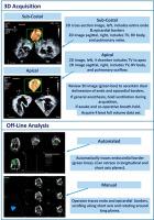
Accurate knowledge of right ventricular (RV) volumes and ejection fraction is fundamental to providing optimal care for pediatric patients with congenital and acquired heart disease, as well as pulmonary hypertension. Traditionally, these volumes have been measured using cardiac magnetic resonance because of its accuracy, reproducibility, and freedom from geometric assumptions. More recently, an increasing number of studies have described the measurement of RV volumes using three-dimensional (3D) echocardiography. In addition, volumes by 3D echocardiography have also been used for outcome research studies in congenital heart surgery. Importantly, 3D echocardiographic acquisitions can be obtained over a small number of cardiac cycles, do not require general anesthesia, and are less costly than CMR. The ease and safety of the 3D echocardiographic acquisitions allow serial studies in the same patient. Moreover, the studies can be performed in various locations, including the intensive care unit, catheterization laboratory, and general clinic. Because of these advantages, 3D echocardiography is ideal for serial evaluation of the same patient. Despite these potential advantages, 3D echocardiography has not become a standard practice in children with congenital and acquired heart conditions. In this report, the authors review the literature on the feasibility, reproducibility, and accuracy of 3D echocardiography in pediatric patients. In addition, the authors investigate the advantages and limitations of 3D echocardiography in RV quantification and offer a pathway for its potential to become a standard practice in the assessment, planning, and follow-up of congenital and acquired heart disease.
中文翻译:

三维超声心动图在小儿心脏病右心室容量分析中的作用的演变:文献综述和临床应用
准确了解右心室 (RV) 容积和射血分数对于为患有先天性和后天性心脏病以及肺动脉高压的儿科患者提供最佳护理至关重要。传统上,这些体积是使用心脏磁共振来测量的,因为它的准确性、可重复性和不受几何假设的影响。最近,越来越多的研究描述了使用三维 (3D) 超声心动图测量右心室容积。此外,3D 超声心动图体积也已用于先天性心脏病手术的结果研究。重要的是,3D 超声心动图采集可以在少量心动周期内获得,不需要全身麻醉,并且比 CMR 成本更低。 3D 超声心动图采集的简便性和安全性允许对同一患者进行系列研究。此外,这些研究可以在不同的地点进行,包括重症监护病房、导管实验室和普通诊所。由于这些优点,3D 超声心动图非常适合对同一患者进行连续评估。尽管有这些潜在优势,3D 超声心动图尚未成为先天性和后天性心脏病儿童的标准治疗方法。在本报告中,作者回顾了有关儿科患者 3D 超声心动图的可行性、可重复性和准确性的文献。此外,作者还研究了 3D 超声心动图在 RV 定量方面的优点和局限性,并为其成为先天性和获得性心脏病的评估、计划和随访标准实践的潜力提供了一条途径。
更新日期:2024-03-11
中文翻译:

三维超声心动图在小儿心脏病右心室容量分析中的作用的演变:文献综述和临床应用
准确了解右心室 (RV) 容积和射血分数对于为患有先天性和后天性心脏病以及肺动脉高压的儿科患者提供最佳护理至关重要。传统上,这些体积是使用心脏磁共振来测量的,因为它的准确性、可重复性和不受几何假设的影响。最近,越来越多的研究描述了使用三维 (3D) 超声心动图测量右心室容积。此外,3D 超声心动图体积也已用于先天性心脏病手术的结果研究。重要的是,3D 超声心动图采集可以在少量心动周期内获得,不需要全身麻醉,并且比 CMR 成本更低。 3D 超声心动图采集的简便性和安全性允许对同一患者进行系列研究。此外,这些研究可以在不同的地点进行,包括重症监护病房、导管实验室和普通诊所。由于这些优点,3D 超声心动图非常适合对同一患者进行连续评估。尽管有这些潜在优势,3D 超声心动图尚未成为先天性和后天性心脏病儿童的标准治疗方法。在本报告中,作者回顾了有关儿科患者 3D 超声心动图的可行性、可重复性和准确性的文献。此外,作者还研究了 3D 超声心动图在 RV 定量方面的优点和局限性,并为其成为先天性和获得性心脏病的评估、计划和随访标准实践的潜力提供了一条途径。


 京公网安备 11010802027423号