European Journal of Nuclear Medicine and Molecular Imaging ( IF 9.1 ) Pub Date : 2024-03-08 , DOI: 10.1007/s00259-024-06670-5 Jennifer Nash , Samuel Debono , Beth Whittington , Jakub Kaczynski , Tim Clark , Gillian Macnaught , Scott Semple , Edwin J R van Beek , Adriana Tavares , Damini Dey , Michelle C Williams , Piotr J Slomka , David E Newby , Marc R Dweck , Alexander J Fletcher
|
Introduction
Non-invasive detection of pathological changes in thoracic aortic disease remains an unmet clinical need particularly for patients with congenital heart disease. Positron emission tomography combined with magnetic resonance imaging (PET-MRI) could provide a valuable low-radiation method of aortic surveillance in high-risk groups. Quantification of aortic microcalcification activity using sodium [18F]fluoride holds promise in the assessment of thoracic aortopathies. We sought to evaluate aortic sodium [18F]fluoride uptake in PET-MRI using three methods of attenuation correction compared to positron emission tomography computed tomography (PET-CT) in patients with bicuspid aortic valve,
Methods
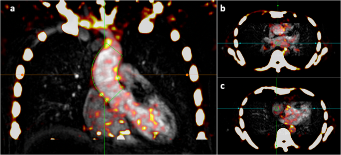
Thirty asymptomatic patients under surveillance for bicuspid aortic valve disease underwent sodium [18F]fluoride PET-CT and PET-MRI of the ascending thoracic aorta during a single visit. PET-MRI data were reconstructed using three iterations of attenuation correction (Dixon, radial gradient recalled echo with two [RadialVIBE-2] or four [RadialVIBE-4] tissue segmentation). Images were qualitatively and quantitatively analysed for aortic sodium [18F]fluoride uptake on PET-CT and PET-MRI.
Results
Aortic sodium [18F]fluoride uptake on PET-MRI was visually comparable with PET-CT using each reconstruction and total aortic standardised uptake values on PET-CT strongly correlated with each PET-MRI attenuation correction method (Dixon R = 0.70; RadialVIBE-2 R = 0.63; RadialVIBE-4 R = 0.64; p < 0.001 for all). Breathing related artefact between soft tissue and lung were detected using Dixon and RadialVIBE-4 but not RadialVIBE-2 reconstructions, with the presence of this artefact adjacent to the atria leading to variations in blood pool activity estimates. Consequently, quantitative agreements between radiotracer activity on PET-CT and PET-MRI were most consistent with RadialVIBE-2.
Conclusion
Ascending aortic microcalcification analysis in PET-MRI is feasible with comparable findings to PET-CT. RadialVIBE-2 tissue attenuation correction correlates best with the reference standard of PET-CT and is less susceptible to artefact. There remain challenges in segmenting tissue types in PET-MRI reconstructions, and improved attenuation correction methods are required.
中文翻译:
正电子发射断层扫描和磁共振联合成像中的胸主动脉微钙化活动
介绍
胸主动脉疾病病理变化的无创检测仍然是一个未满足的临床需求,特别是对于先天性心脏病患者。正电子发射断层扫描与磁共振成像(PET-MRI)相结合可以为高危人群的主动脉监测提供一种有价值的低辐射方法。使用[ 18 F]氟化钠定量主动脉微钙化活性在胸主动脉病的评估中具有前景。我们试图使用三种衰减校正方法与二叶式主动脉瓣患者的正电子发射断层扫描计算机断层扫描 (PET-CT) 相比,评估 PET-MRI 中主动脉钠[ 18 F]氟化物的摄取,
方法
30 名接受二叶式主动脉瓣疾病监测的无症状患者在单次就诊期间接受了[ 18 F]氟化钠 PET-CT 和升胸主动脉 PET-MRI。使用三次衰减校正迭代重建 PET-MRI 数据(Dixon、使用两个 [RadialVIBE-2] 或四个 [RadialVIBE-4] 组织分割的径向梯度回忆回波)。对PET-CT 和 PET-MRI 上的主动脉[ 18 F]氟化钠摄取图像进行定性和定量分析。
结果
PET-MRI 上的主动脉钠[ 18 F]氟化物摄取在视觉上与 PET-CT 具有可比性,使用每次重建和 PET-CT 上的总主动脉标准化摄取值与每种 PET-MRI 衰减校正方法密切相关(Dixon R = 0.70;RadialVIBE- 2 R = 0.63;RadialVIBE-4 R = 0.64;p < 0.001)。使用 Dixon 和 RadialVIBE-4(而非 RadialVIBE-2 重建)检测到软组织和肺部之间与呼吸相关的伪影,这种伪影靠近心房的存在导致血池活动估计的变化。因此,PET-CT 和 PET-MRI 上的放射性示踪剂活性之间的定量一致性与 RadialVIBE-2 最为一致。
结论
PET-MRI 中的升主动脉微钙化分析是可行的,其结果与 PET-CT 相当。RadialVIBE-2 组织衰减校正与 PET-CT 参考标准的相关性最好,并且不易受到伪影的影响。PET-MRI 重建中的组织类型分割仍然存在挑战,需要改进的衰减校正方法。


 京公网安备 11010802027423号