当前位置:
X-MOL 学术
›
Phys. Rev. X
›
论文详情
Our official English website, www.x-mol.net, welcomes your
feedback! (Note: you will need to create a separate account there.)
Modeling and Predicting Second-Harmonic Generation from Protein Molecular Structure
Physical Review X ( IF 11.6 ) Pub Date : 2024-03-06 , DOI: 10.1103/physrevx.14.011038 Bahar Asadipour , Emmanuel Beaurepaire , Xingjian Zhang , Anatole Chessel , Pierre Mahou , Willy Supatto , Marie-Claire Schanne-Klein , Chiara Stringari
Physical Review X ( IF 11.6 ) Pub Date : 2024-03-06 , DOI: 10.1103/physrevx.14.011038 Bahar Asadipour , Emmanuel Beaurepaire , Xingjian Zhang , Anatole Chessel , Pierre Mahou , Willy Supatto , Marie-Claire Schanne-Klein , Chiara Stringari
|
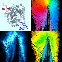
Polarization resolved second-harmonic generation (pSHG) microscopy is increasingly used for mapping organized arrays of noncentrosymmetric proteins such as collagen, myosin, and tubulin, and holds potential for probing their molecular structure and supramolecular organization in intact tissues. However, the contrast mechanism of pSHG is complex and the development of applications in the life sciences is hampered by the lack of models accurately relating the observed pSHG signals to the underlying molecular and macromolecular organization. In this work, we establish a general multiscale numerical framework relating the micrometer-scale SHG measurements to the atomic-scale and molecular structure of the proteins under study and their supramolecular arrangement. We first develop a new method to automatically analyze pSHG signals independently of the protein type and fiber orientation. We then characterize experimentally pSHG signals in live zebrafish larvae and show that they can be used to distinguish collagen, myosin, and tubulin structures in intact tissues. We then introduce a numerical model that considers the peptide bond (PB) as the elementary SHG source in proteins and takes into account the three-dimensional (3D) distribution of PBs to predict the second-order hyperpolarizability tensor of proteins, as well as the SHG efficiency and pSHG response of an arbitrary macromolecular assembly. We show that this model accurately reproduces pSHG measurements obtained from collagen, myosin, microtubule, and actin structures, revealing the precise dependence of SHG signals on the 3D distribution of PBs within protein assemblies. We then use our model to analyze pSHG from a 3D distribution of microtubule assemblies as a function of out-of-plane angles, angular disorder, and polarity. Finally, we demonstrate that our model predicts SHG from different molecular conformations of tubulin that are highly relevant from a biomedical point of view as associated with microtubules (de)polymerization. By bridging scales from the molecular bonds to the optical wavelength, our model provides an accurate interpretation of SHG signals in terms of protein structure and supramolecular organization.
中文翻译:

根据蛋白质分子结构建模和预测二次谐波的产生
偏振分辨二次谐波发生 (pSHG) 显微镜越来越多地用于绘制胶原蛋白、肌球蛋白和微管蛋白等非中心对称蛋白质的有序阵列图谱,并具有探测完整组织中其分子结构和超分子组织的潜力。然而,pSHG 的对比机制很复杂,并且由于缺乏将观察到的 pSHG 信号与底层分子和大分子组织准确关联的模型,阻碍了其在生命科学中的应用发展。在这项工作中,我们建立了一个通用的多尺度数值框架,将微米级二次谐波测量与所研究蛋白质的原子尺度和分子结构及其超分子排列联系起来。我们首先开发了一种新方法来自动分析 pSHG 信号,而与蛋白质类型和纤维方向无关。然后,我们通过实验表征活体斑马鱼幼虫中的 pSHG 信号,并表明它们可用于区分完整组织中的胶原蛋白、肌球蛋白和微管蛋白结构。然后,我们引入了一个数值模型,该模型将肽键 (PB) 视为蛋白质中的基本 SHG 源,并考虑 PB 的三维 (3D) 分布来预测二阶超极化张量蛋白质的分析,以及任意大分子组装体的 SHG 效率和 pSHG 响应。我们表明,该模型准确地再现了从胶原蛋白、肌球蛋白、微管和肌动蛋白结构中获得的 pSHG 测量结果,揭示了 SHG 信号对蛋白质组装体中 PB 3D 分布的精确依赖性。然后,我们使用我们的模型从微管组件的 3D 分布中分析 pSHG 作为面外角度、角度无序和极性的函数。最后,我们证明我们的模型可以根据微管蛋白的不同分子构象预测 SHG,从生物医学的角度来看,这些构象与微管(解)聚合高度相关。通过桥接分子键和光波长的尺度,我们的模型可以在蛋白质结构和超分子组织方面准确解释 SHG 信号。
更新日期:2024-03-06
中文翻译:
根据蛋白质分子结构建模和预测二次谐波的产生
偏振分辨二次谐波发生 (pSHG) 显微镜越来越多地用于绘制胶原蛋白、肌球蛋白和微管蛋白等非中心对称蛋白质的有序阵列图谱,并具有探测完整组织中其分子结构和超分子组织的潜力。然而,pSHG 的对比机制很复杂,并且由于缺乏将观察到的 pSHG 信号与底层分子和大分子组织准确关联的模型,阻碍了其在生命科学中的应用发展。在这项工作中,我们建立了一个通用的多尺度数值框架,将微米级二次谐波测量与所研究蛋白质的原子尺度和分子结构及其超分子排列联系起来。我们首先开发了一种新方法来自动分析 pSHG 信号,而与蛋白质类型和纤维方向无关。然后,我们通过实验表征活体斑马鱼幼虫中的 pSHG 信号,并表明它们可用于区分完整组织中的胶原蛋白、肌球蛋白和微管蛋白结构。然后,我们引入了一个数值模型,该模型将肽键 (PB) 视为蛋白质中的基本 SHG 源,并考虑 PB 的三维 (3D) 分布来预测二阶超极化张量蛋白质的分析,以及任意大分子组装体的 SHG 效率和 pSHG 响应。我们表明,该模型准确地再现了从胶原蛋白、肌球蛋白、微管和肌动蛋白结构中获得的 pSHG 测量结果,揭示了 SHG 信号对蛋白质组装体中 PB 3D 分布的精确依赖性。然后,我们使用我们的模型从微管组件的 3D 分布中分析 pSHG 作为面外角度、角度无序和极性的函数。最后,我们证明我们的模型可以根据微管蛋白的不同分子构象预测 SHG,从生物医学的角度来看,这些构象与微管(解)聚合高度相关。通过桥接分子键和光波长的尺度,我们的模型可以在蛋白质结构和超分子组织方面准确解释 SHG 信号。


 京公网安备 11010802027423号