The Pharmacogenomics Journal ( IF 2.8 ) Pub Date : 2024-02-20 , DOI: 10.1038/s41397-024-00324-3 Peng-Wei Zhao , Jia-Xian Cui , Xiu-Mei Wang
|
Objective
To explore the role of p300 in the context of paclitaxel (PTX) resistance in triple-negative breast cancer (TNBC) cells, focusing on its interaction with the phosphoenolpyruvate carboxykinase 1 (PCK1)/adenosine monophosphate-activated protein kinase (AMPK) pathway.
Methods
The expression of p300 and PCK1 at the messenger ribonucleic acid (mRNA) level was detected using a quantitative polymerase chain reaction. The GeneCards and GEPIA databases were used to investigate the relationship between p300 and PCK1. The MDA-MB-231/PTX cell line, known for its PTX resistance, was chosen to understand the specific role of p300 in such cells. The Lipofectamine™ 3000 reagent was used to transfer the p300 small interfering RNA and the overexpression of PCK1 plasmid into MDA-MB-231/PTX. The expression levels of p300, PCK1, 5′AMPK and phosphorylated AMPK (p-AMPK) were determined using the western blot test.
Results
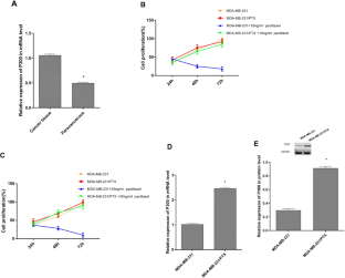
In TNBC cancer tissue, the expression of p300 was increased compared with TNBC paracancerous tissue (P < 0.05). In the MDA-MB-231 cell line of TNBC, the expression of p300 was lower than in the PTX-resistant TNBC cells (MDA-MB-231/PTX) (P < 0.05). The PCK1 expression was decreased in the TNBC cancer tissue compared with TNBC paracancerous tissue, and the PCK1 expression was reduced in MDA-MB-231/PTX than in MDA-MB-231 (P < 0.05) indicating that PCK1 was involved in the resistance function. Additionally, p-AMPK was decreased in MDA-MB-231/PTX compared with MDA-MB-231 (P < 0.05). The adenosine triphosphate (ATP) level was also detected and was significantly lower in MDA-MB-231/PTX than in MDA-MB-231 (P < 0.05). Additionally, cell proliferation increased significantly in MDA-MB-231/PTX at 48 and 72 h (P < 0.05) suggesting that MDA-MB-231/PTX cells obtained the resistance function which was associated with AMPK and ATP level. When p300 was inhibited, p-AMPK and ATP levels elevated in MDA-MB-231/PTX (P < 0.05). When PCK1 was suppressed, the ATP consumption rate decreased, and cell proliferation increased (P < 0.05). However, there were no changes in p300.
Conclusions
In MDA-MB-231/PTX, p300 can inhibit p-AMPK and ATP levels by inhibiting PCK1 expression. Our findings suggest that targeting p300 could modulate the PCK1/AMPK axis, offering a potential therapeutic avenue for overcoming PTX resistance in TNBC.
中文翻译:
紫杉醇耐药 TNBC 中 p300 的上调:通过 PCK1/AMPK 轴对细胞增殖的影响
客观的
探讨 p300 在三阴性乳腺癌 (TNBC) 细胞紫杉醇 (PTX) 耐药中的作用,重点关注其与磷酸烯醇丙酮酸羧激酶 1 (PCK1)/腺苷单磷酸激活蛋白激酶 (AMPK) 通路的相互作用。
方法
使用定量聚合酶链式反应检测信使核糖核酸 (mRNA) 水平的 p300 和 PCK1 表达。GeneCards 和 GEPIA 数据库用于研究 p300 和 PCK1 之间的关系。选择以其 PTX 耐药性而闻名的 MDA-MB-231/PTX 细胞系来了解 p300 在此类细胞中的具体作用。使用 Lipofectamine™ 3000 试剂将 p300 小干扰 RNA 和 PCK1 质粒的过表达转移至 MDA-MB-231/PTX 中。使用蛋白质印迹测试测定p300、PCK1、5'AMPK和磷酸化AMPK(p-AMPK)的表达水平。
结果
TNBC 癌组织中 p300 表达量较 TNBC 癌旁组织升高(P < 0.05)。TNBC MDA-MB-231 细胞系中 p300 的表达低于 PTX 耐药 TNBC 细胞(MDA-MB-231/PTX)(P < 0.05)。与 TNBC 癌旁组织相比,TNBC 癌组织中 PCK1 表达量降低,且 MDA-MB-231/PTX 中 PCK1 表达量较 MDA-MB-231 降低(P < 0.05),表明 PCK1 参与了耐药。功能。此外,与 MDA-MB-231 相比,MDA-MB-231/PTX 中的 p-AMPK 降低(P < 0.05)。还检测到三磷酸腺苷 (ATP) 水平,MDA-MB-231/PTX 中的三磷酸腺苷 (ATP) 水平显着低于 MDA-MB-231 ( P < 0.05)。此外,MDA-MB-231/PTX在48和72 h时细胞增殖显着增加(P < 0.05),表明MDA-MB-231/PTX细胞获得了与AMPK和ATP水平相关的耐药功能。当 p300 受到抑制时,MDA-MB-231/PTX 中的 p-AMPK 和 ATP 水平升高(P < 0.05)。当 PCK1 受到抑制时,ATP 消耗率降低,细胞增殖增加(P < 0.05)。然而,p300 没有变化。
结论
在MDA-MB-231/PTX中,p300可以通过抑制PCK1表达来抑制p-AMPK和ATP水平。我们的研究结果表明,靶向 p300 可以调节 PCK1/AMPK 轴,为克服 TNBC 中的 PTX 耐药性提供潜在的治疗途径。


 京公网安备 11010802027423号