Nature Photonics ( IF 35.0 ) Pub Date : 2024-02-16 , DOI: 10.1038/s41566-024-01387-1 Youjuan Wang , Zhigao Yi , Jing Guo , Shiyi Liao , Zhe Li , Shuai Xu , Baoli Yin , Yongchao Liu , Yurong Feng , Qiming Rong , Xiaogang Liu , Guosheng Song , Xiao-Bing Zhang , Weihong Tan
|
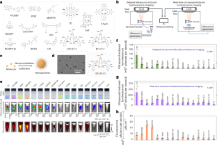
Optical imaging is crucial to study biological or pathological processes and diagnose diseases. However, poor tissue penetration typically limits conventional optical imaging. Here we report an imaging technique that uses ultrasound to activate luminescent molecules or nanoparticles through two-step intraparticle energy conversion. Ultrasonic activation can convert mechanical fluctuations into chemical energy via the piezoelectric effect and then induce luminescence via the chemiluminescent effect. We demonstrate two modalities for ultrasound-induced luminescence imaging: one achieves delayed imaging after cessation of the ultrasonic excitation, and the other enables real-time imaging during the ultrasonic excitation. Our imaging modality offers an improvement in luminescence intensity of up to 2,000-fold compared with sonoluminescence of H2O, a 10-fold improved of signal-to-noise ratio compared with fluorescence imaging, a spatial resolution of 1.46 mm and tissue penetration of up to 2.2 cm. We demonstrate its applicability for imaging subcutaneous and orthotopic tumours, mapping lymph nodes and screening peritoneal metastatic tumours. Furthermore, we design analyte-activatable luminescence probes based on resonance energy transfer, which can assess drug-induced hepatotoxicity and distinguish the responsivity of tumours after drug treatment. We expect that our technique will enable further preclinical and clinical applications, such as the study of histopathological lesions in living animals, the early detection of tumours, the profiling of biological molecules and the monitoring of cancer treatment or prognosis, among others.
中文翻译:
体内超声诱导发光分子成像
光学成像对于研究生物或病理过程以及诊断疾病至关重要。然而,较差的组织穿透力通常限制了传统的光学成像。在这里,我们报告了一种成像技术,该技术使用超声波通过两步粒子内能量转换来激活发光分子或纳米粒子。超声波激活可以通过压电效应将机械波动转化为化学能,然后通过化学发光效应诱导发光。我们展示了超声诱导发光成像的两种模式:一种在超声激励停止后实现延迟成像,另一种在超声激励期间实现实时成像。我们的成像方式与 H 2 O声致发光相比,发光强度提高了 2,000 倍,与荧光成像相比,信噪比提高了 10 倍,空间分辨率为 1.46 mm,组织穿透力为最大 2.2 厘米。我们证明了它在皮下和原位肿瘤成像、淋巴结定位和筛查腹膜转移肿瘤方面的适用性。此外,我们还设计了基于共振能量转移的可分析物激活的发光探针,可以评估药物引起的肝毒性并区分药物治疗后肿瘤的反应性。我们期望我们的技术能够进一步实现临床前和临床应用,例如活体动物组织病理学病变的研究、肿瘤的早期检测、生物分子的分析以及癌症治疗或预后的监测等。


 京公网安备 11010802027423号