European Journal of Nuclear Medicine and Molecular Imaging ( IF 9.1 ) Pub Date : 2024-02-13 , DOI: 10.1007/s00259-024-06650-9 Konstantinos G. Zeimpekis , Lorenzo Mercolli , Maurizio Conti , Hasan Sari , Axel Rominger , Hendrik Rathke
|
Purpose
Evaluation of 90Y liver radioembolization post-treatment clinical data using a whole-body Biograph Vision Quadra PET/CT to investigate the potential of protocol optimization in terms of scan time and dosimetry.
Methods
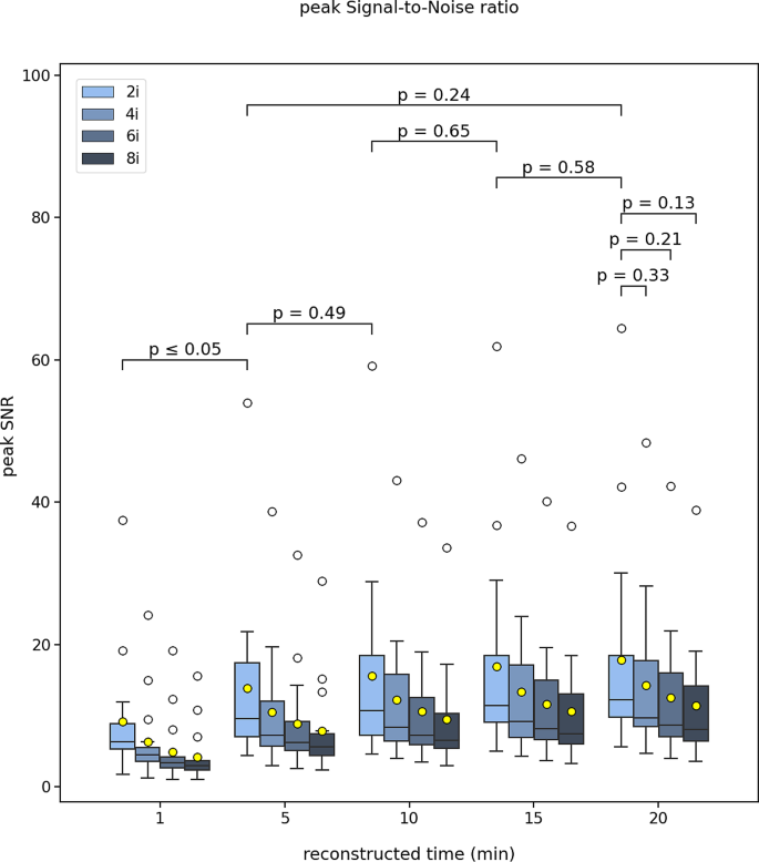
17 patients with hepatocellular carcinoma with median (IQR) injected activity 2393 (1348–3298) MBq were included. Pre-treatment dosimetry plan was based on 99mTc-MAA SPECT/CT with Simplicit90Y™ and post-treatment validation with Quadra using Simplicit90Y™ and HERMIA independently. Regarding the image analysis, mean and peak SNR, the coefficient of variation (COV) and lesion-to-background ratio (LBR) were evaluated. For the post-treatment dosimetry validation, the mean tumor, whole liver and lung absorbed dose evaluation was performed using Simplicit90Y and HERMES. Images were reconstructed with 20-, 15-, 10-, 5- and 1- min sinograms with 2, 4, 6 and 8 iterations. Wilcoxon signed rank test was used to show statistical significance (p < 0.05).
Results
There was no difference of statistical significance between 20- and 5- min reconstructed times for the peak SNR, COV and LBR. In addition, there was no difference of statistical significance between 20- and 1- min reconstructed times for all dosimetry metrics. Lung dosimetry showed consistently lower values than the expected. Tumor absorbed dose based on Simplicit90Y™ was similar to the expected while HERMES consistently underestimated significantly the measured tumor absorbed dose. Finally, there was no difference of statistical significance between expected and measured tumor, whole liver and lung dose for all reconstruction times.
Conclusion
In this study we evaluated, in terms of image quality and dosimetry, whole-body PET clinical images of patients after having been treated with 90Y microspheres radioembolization for liver cancer. Compared to the 20-min standard scan, the simulated 5-min reconstructed images provided equal image peak SNR and noise behavior, while performing also similarly for post-treatment dosimetry of tumor, whole liver and lung absorbed doses.


 京公网安备 11010802027423号