npj Microgravity ( IF 5.1 ) Pub Date : 2024-01-17 , DOI: 10.1038/s41526-024-00347-x Yuan Xie , Yingdi Fu , Yaqi Shao , Lina Qu , Jiangang Yang , Chengjia Yang , Kun Zhou , Kai Li , Zi Xu , Dong Xu , Kai Cao , Ning Tian , Ke Lv , Linjie Wang , Yaping Wang , Ningli Wang , Yinghui Li
|
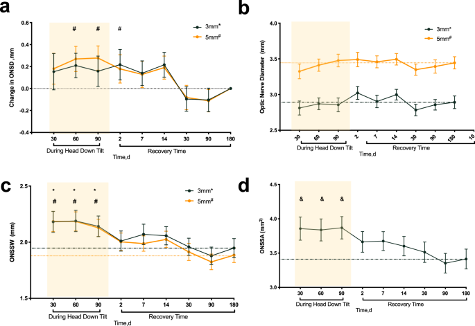
The elevation in the optic nerve sheath (ONS) pressure (ONSP) due to microgravity-induced headward fluid shift is the primary hypothesized contributor to SANS. This longitudinal study aims to quantify the axial plane of the optic nerve subarachnoid space area (ONSSA), which is filled with cerebrospinal fluid (CSF) and expands with elevated ONSP during and after head-down tilt (HDT) bed rest (BR). 36 healthy male volunteers (72 eyes) underwent a 90-day strict 6° HDT BR. Without obtaining the pre-HDT data, measurements were performed on days 30, 60, and 90 during HDT and at 6 recovery time points extended to 180-days (R + 180) in a supine position. Portable B-scan ultrasound was performed using the 12 MHz linear array probe binocularly. The measurements of the ONS and the calculation of the ONSSA were performed with ImageJ 1.51 analysis software by two experienced observers in a masked manner. Compared to R + 180, the ONSSA on HDT30, HDT60, and HDT90 exhibited a consistently significant distention of 0.44 mm2 (95% CI: 0.13 to 0.76 mm2, P = 0.001), 0.45 mm2 (95% CI: 0.15 to 0.75 mm2, P = 0.001), and 0.46 mm2 (95% CI: 0.15 to 0.76 mm2, P < 0.001), respectively, and recovered immediately after HDT on R + 2. Such small changes in the ONSSA were below the lateral resolution limit of ultrasound (0.4 mm) and may not be clinically relevant, possibly due to ONS hysteresis causing persistent ONS distension. Future research can explore advanced quantitative portable ultrasound-based techniques and establish comparisons containing the pre-HDT measurements to deepen our understanding of SANS.
中文翻译:
90天头低位倾斜卧床休息期间视神经蛛网膜下腔的定量超声图像评估
由于微重力引起的液体向头移动而引起的视神经鞘 (ONS) 压力 (ONSP) 升高是 SANS 的主要假设因素。这项纵向研究旨在量化视神经蛛网膜下腔区域 (ONSSA) 的轴平面,该区域充满脑脊液 (CSF),并在低头倾斜 (HDT) 卧床休息 (BR) 期间和之后随着 ONSP 的升高而扩张。36 名健康男性志愿者(72 只眼睛)接受了为期 90 天的严格 6° HDT BR。在没有获得 HDT 前数据的情况下,在 HDT 期间的第 30、60 和 90 天以及延长至 180 天(R + 180)的 6 个恢复时间点以仰卧位进行测量。使用 12 MHz 线性阵列探头双目进行便携式 B 扫描超声检查。ONS 的测量和 ONSSA 的计算由两名经验丰富的观察员以屏蔽方式使用 ImageJ 1.51 分析软件进行。与 R + 180 相比,HDT30、HDT60 和 HDT90 上的 ONSSA 表现出一致的显着膨胀,分别为 0.44 mm 2(95% CI:0.13 至 0.76 mm 2,P = 0.001)、0.45 mm 2(95% CI:0.15 至分别为 0.75 mm 2,P = 0.001) 和 0.46 mm 2 (95% CI:0.15 至 0.76 mm 2,P < 0.001),并且在 R + 2 上的 HDT 后立即恢复。ONSSA 的如此小的变化低于超声的横向分辨率限制(0.4 毫米),可能与临床无关,可能是由于 ONS 滞后导致持续 ONS 扩张。未来的研究可以探索先进的定量便携式超声技术,并建立包含 HDT 前测量值的比较,以加深我们对 SANS 的理解。


 京公网安备 11010802027423号