Progress in Retinal and Eye Research ( IF 17.8 ) Pub Date : 2023-02-12 , DOI: 10.1016/j.preteyeres.2023.101170 Grazyna Palczewska 1 , Maciej Wojtkowski 2 , Krzysztof Palczewski 3
|
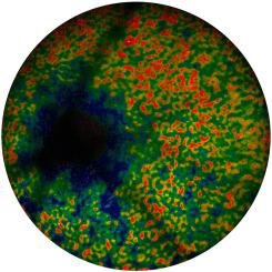
The eye is an ideal organ for imaging by a multi-photon excitation approach, because ocular tissues such as the sclera, cornea, lens and neurosensory retina, are highly transparent to infrared (IR) light. The interface between the retina and the retinal pigment epithelium (RPE) is especially informative, because it reflects the health of the visual (retinoid) cycle and its changes in response to external stress, genetic manipulations, and drug treatments. Vitamin A-derived retinoids, like retinyl esters, are natural fluorophores that respond to multi-photon excitation with near IR light, bypassing the filter-like properties of the cornea, lens, and macular pigments. Also, during natural aging some retinoids form bisretinoids, like diretinoid-pyridiniumethanolamine (A2E), that are highly fluorescent. These bisretinoids appear to be elevated concurrently with aging. Vitamin A-derived retinoids and bisretinoidss are detected by two-photon ophthalmoscopy (2PO), using a new class of light sources with adjustable spatial, temporal, and spectral properties. Furthermore, the two-photon (2P) absorption of IR light by the visual pigments in rod and cone photoreceptors can initiate visual transduction by cis-trans isomerization of retinal, enabling parallel functional studies. Recently we overcame concerns about safety, data interpretation and complexity of the 2P-based instrumentation, the major roadblocks toward advancing this modality to the clinic. These imaging and retina-function assessment advancements have enabled us to conduct the first 2P studies with humans.
中文翻译:
从小鼠到人类:通过双光子激发了解体内视觉的生物化学
眼睛是通过多光子激发方法成像的理想器官,因为巩膜、角膜、晶状体和神经感觉视网膜等眼组织对红外 (IR) 光高度透明。视网膜和视网膜色素上皮 (RPE) 之间的界面尤其能提供丰富的信息,因为它反映了视觉(类维生素A)周期的健康状况及其对外部压力、基因操作和药物治疗的反应的变化。维生素 A 衍生的类视黄醇与视黄酯一样,是天然荧光团,可响应近红外光的多光子激发,绕过角膜、晶状体和黄斑色素的类似滤光片的特性。此外,在自然老化过程中,一些类维生素A会形成双类维生素A,例如二类维生素A-吡啶乙醇胺(A2E),它们具有高荧光。这些双维A酸似乎随着衰老而同时升高。维生素 A 衍生的类视黄醇和双视黄醇通过双光子检眼镜 (2PO) 进行检测,使用具有可调节空间、时间和光谱特性的新型光源。此外,视杆细胞和视锥细胞光感受器中的视觉色素对红外光的双光子(2P)吸收可以通过视网膜的顺反异构化启动视觉转导,从而实现并行功能研究。最近,我们克服了对基于 2P 的仪器的安全性、数据解释和复杂性的担忧,这些是将该模式推向临床的主要障碍。这些成像和视网膜功能评估的进步使我们能够对人类进行首次 2P 研究。


 京公网安备 11010802027423号