当前位置:
X-MOL 学术
›
J. Am. Chem. Soc.
›
论文详情
Our official English website, www.x-mol.net, welcomes your
feedback! (Note: you will need to create a separate account there.)
Mitochondrial Imaging with Combined Fluorescence and Stimulated Raman Scattering Microscopy Using a Probe of the Aggregation-Induced Emission Characteristic
Journal of the American Chemical Society ( IF 14.4 ) Pub Date : 2017-11-16 00:00:00 , DOI: 10.1021/jacs.7b06273 Xuesong Li 1 , Meijuan Jiang 1 , Jacky W. Y. Lam 1 , Ben Zhong Tang 1 , Jianan Y. Qu 1
Journal of the American Chemical Society ( IF 14.4 ) Pub Date : 2017-11-16 00:00:00 , DOI: 10.1021/jacs.7b06273 Xuesong Li 1 , Meijuan Jiang 1 , Jacky W. Y. Lam 1 , Ben Zhong Tang 1 , Jianan Y. Qu 1
Affiliation

|
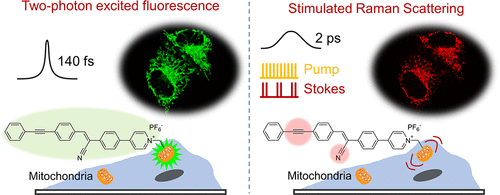
In vivo quantitative measurement of biodistribution plays a critical role in the drug/probe development and diagnosis/treatment process monitoring. In this work, we report a probe, named AIE-SRS-Mito, for imaging mitochondria in live cells via fluorescence (FL) and stimulated Raman scattering (SRS) imaging. The probe features an aggregation-induced emission (AIE) characteristic and possesses an enhanced alkyne Raman peak at 2223 cm–1. The dual-mode imaging of AIE-SRS-Mito for selective mitochondrion-targeting was examined on a homemade FL–SRS microscope system. The detection limit of the probe in the SRS imaging was estimated to be 8.5 μM. Due to the linear concentration dependence of SRS and inertness of the alkyne Raman signal to environmental changes, the intracellular distribution of the probe was studied, showing a local concentration of >2.0 mM in the mitochondria matrix, which was >100-fold higher than the incubation concentration. To the best of our knowledge, this is the first time that the local concentration of AIE molecules inside cells has been measured noninvasively and directly. Also, the nonquenching effect of such AIE molecules in cell imaging has been verified by the positive correlation of FL and SRS signals. Our work will encourage the utilization of SRS microscopy for quantitative characterization of FL probes or other nonfluorescent compounds in living biological systems and the development of FL–SRS dual-mode probes for specific biotargets.
中文翻译:

线粒体成像结合荧光和受激拉曼散射显微镜使用聚集诱导发射特征的探针。
体内生物分布的定量测量在药物/探针开发以及诊断/治疗过程监测中起着至关重要的作用。在这项工作中,我们报告了一个名为AIE-SRS-Mito的探针,用于通过荧光(FL)和受激拉曼散射(SRS)成像对活细胞中的线粒体进行成像。该探针具有聚集诱导发射(AIE)特征,并在2223 cm –1处具有增强的炔烃拉曼峰。在自制的FL-SRS显微镜系统上检查了用于选择性线粒体靶向的AIE-SRS-Mito的双模成像。SRS成像中探针的检出限估计为8.5μM。由于SRS的线性浓度依赖性以及炔烃拉曼信号对环境变化的惰性,因此研究了探针的细胞内分布,结果表明线粒体基质中的局部浓度> 2.0 mM,比线粒体基质的浓度高100倍以上。孵育浓度。据我们所知,这是首次无创,直接地测量细胞内AIE分子的局部浓度。而且,通过FL和SRS信号的正相关已经证实了这种AIE分子在细胞成像中的非猝灭作用。
更新日期:2017-11-17
中文翻译:

线粒体成像结合荧光和受激拉曼散射显微镜使用聚集诱导发射特征的探针。
体内生物分布的定量测量在药物/探针开发以及诊断/治疗过程监测中起着至关重要的作用。在这项工作中,我们报告了一个名为AIE-SRS-Mito的探针,用于通过荧光(FL)和受激拉曼散射(SRS)成像对活细胞中的线粒体进行成像。该探针具有聚集诱导发射(AIE)特征,并在2223 cm –1处具有增强的炔烃拉曼峰。在自制的FL-SRS显微镜系统上检查了用于选择性线粒体靶向的AIE-SRS-Mito的双模成像。SRS成像中探针的检出限估计为8.5μM。由于SRS的线性浓度依赖性以及炔烃拉曼信号对环境变化的惰性,因此研究了探针的细胞内分布,结果表明线粒体基质中的局部浓度> 2.0 mM,比线粒体基质的浓度高100倍以上。孵育浓度。据我们所知,这是首次无创,直接地测量细胞内AIE分子的局部浓度。而且,通过FL和SRS信号的正相关已经证实了这种AIE分子在细胞成像中的非猝灭作用。


 京公网安备 11010802027423号